The KTN1 Antibody (CAB12456) is a high-quality antibody developed for reliable detection and analysis of target proteins. This antibody, derived from rabbit serum, shows high specificity and sensitivity towards KTN1 in human samples, making it an ideal choice for Western blot applications. By targeting the KTN1 protein, this antibody enables accurate detection and analysis in various cell types, allowing for in-depth studies in areas such as cell biology and cancer research.KTN1, also known as kinectin, is essential for proper cell division and organization of the cytoskeleton, making it a crucial protein for maintaining cellular function and structure.
This antibody is validated for use in WB, ELISA applications and has demonstrated reactivity against Human, Mouse, Rat samples.
Product Name:
KTN1 Antibody
SKU:
CAB12456
Size:
20μL, 100μL
Reactivity:
Human, Mouse, Rat
Conjugate:
Unconjugated
Immunogen:
Synthetic peptide. This information is considered to be commercially sensitive.
Recommended starting concentration is 1 μg/mL. Please optimize the concentration based on your specific assay requirements.
Synonyms:
CG1, KNT, MU-RMS-40.19, KTN1
Positive Sample:
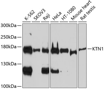
K-562, SKOV3, Raji, HeLa, HT-1080, Mouse heart, Rat testis
Cellular Localization:
Endoplasmic Reticulum Membrane, Single-Pass Type Ii Membrane Protein.
Calculated MW:
156kDa
Observed MW:
178kDa
This gene encodes an integral membrane protein that is a member of the kinectin protein family. The encoded protein is primarily localized to the endoplasmic reticulum membrane. This protein binds kinesin and may be involved in intracellular organelle motility. This protein also binds translation elongation factor-delta and may be involved in the assembly of the elongation factor-1 complex. Alternate splicing results in multiple transcript variants of this gene.
Purification Method
Affinity purification
Gene ID
3895
RRID
AB_2759300
Buffer Information
Store at -20℃. Avoid freeze / thaw cycles. Buffer: PBS containing 50% glycerol, preserved with proclin300 or sodium azide, pH 7.3.
Western blot analysis of various lysates using KTN1 Rabbit pAb (CAB12456) at 1:1000 dilution. Secondary antibody: HRP-conjugated Goat anti-Rabbit IgG (H+L) (CABS014) at 1:10000 dilution. Lysates/proteins: 25μg per lane. Blocking buffer: 3% nonfat dry milk in TBST. Detection: ECL Basic Kit (AbGn00020). Exposure time: 90s.