The LAMP1 Antibody (CAB16894) is a high-quality antibody developed for reliable detection and analysis of target proteins. LAMP1 is a key marker for lysosomal compartments and plays a crucial role in regulating lysosomal biogenesis and function. This antibody, generated in rabbits, is highly specific to human samples and has been validated for use in Western blot and immunofluorescence applications.LAMP1 is essential for maintaining cellular homeostasis by mediating lysosomal fusion and autophagosome-lysosome fusion, processes that are vital for degradation of cellular waste and recycling of cellular components. Dysregulation of LAMP1 has been linked to various diseases, including lysosomal storage disorders, neurodegenerative diseases, and cancer.
This antibody is validated for use in WB, IHC-P, IF/ICC, ELISA applications and has demonstrated reactivity against Human, Mouse, Rat samples.
Product Name:
LAMP1 Antibody
SKU:
CAB16894
Size:
20μL, 100μL
Reactivity:
Human, Mouse, Rat
Conjugate:
Unconjugated
Immunogen:
Synthetic peptide. This information is considered to be commercially sensitive.
Recommended starting concentration is 1 μg/mL. Please optimize the concentration based on your specific assay requirements.
Synonyms:
LAMPA, CD107a, LGP120, LAMP1
Positive Sample:
HeLa
Cellular Localization:
Cell Membrane, Endosome Membrane, Late Endosome, Lysosome Membrane, Single-Pass Type I Membrane Protein.
Calculated MW:
45kDa
Observed MW:
42kDa/90-120kDa
The protein encoded by this gene is a member of a family of membrane glycoproteins. This glycoprotein provides selectins with carbohydrate ligands. It may also play a role in tumor cell metastasis.
Purification Method
Affinity purification
Gene ID
3916
RRID
AB_2770145
Buffer Information
Store at -20℃. Avoid freeze / thaw cycles. Buffer: PBS with 0.09% Sodium azide,50% glycerol,pH7.3.
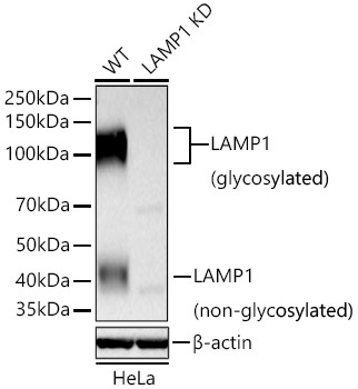
Western blot analysis of lysates from wild type (WT) and LAMP1 knockdown (KD) HeLa cells using [KD Validated] LAMP1 Rabbit pAb (CAB16894) at 1:2000 dilution incubated overnight at 4℃. Secondary antibody: HRP-conjugated Goat anti-Rabbit IgG (H+L) (CABS014) at 1:10000 dilution. Lysates/proteins: 25 μg per lane. Blocking buffer: 3% nonfat dry milk in TBST. Detection: ECL Basic Kit (AbGn00020). Exposure time: 90s.
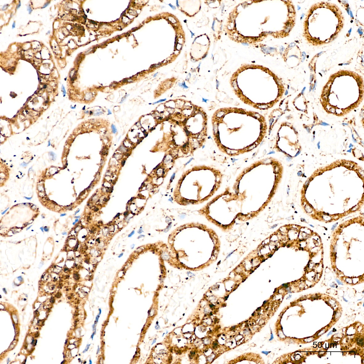
Immunohistochemistry analysis of paraffin-embedded Human kidney tissue using LAMP1 Rabbit pAb (CAB16894) at a dilution of 1:100 (40x lens). High pressure antigen retrieval performed with 0.01M Citrate buffer (pH 6.0) prior to IHC staining.
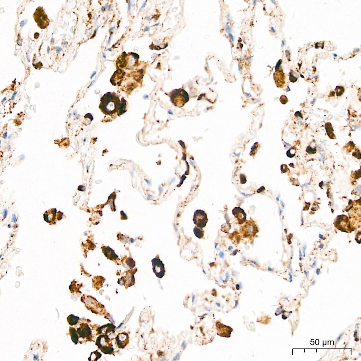
Immunohistochemistry analysis of paraffin-embedded Human lung tissue using LAMP1 Rabbit pAb (CAB16894) at a dilution of 1:100 (40x lens). High pressure antigen retrieval performed with 0.01M Citrate buffer (pH 6.0) prior to IHC staining.
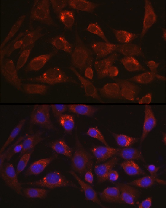
Immunofluorescence analysis of BALA-3T3 cells using LAMP1 Rabbit pAb (CAB16894) at dilution of 1:100 (40x lens). Secondary antibody: Cy3-conjugated Goat anti-Rabbit IgG (H+L) (CABS007) at 1:500 dilution. Blue: DAPI for nuclear staining.