The LAMP3 Antibody (CAB2895) is a high-quality antibody developed for reliable detection and analysis of target proteins. This polyclonal antibody, raised in rabbits, is highly reactive with human samples and is validated for use in various applications, including Western blotting.LAMP3, also known as CD208, is a key player in the process of antigen presentation and immune responses. It is involved in the regulation of autophagy and plays a role in the immune response to pathogens. The LAMP3 Polyclonal Antibody specifically binds to the LAMP3 protein, allowing for the detection and analysis of LAMP3 expression in different cell types.
This antibody is validated for use in WB, IHC-P, ELISA applications and has demonstrated reactivity against Human, Mouse, Rat samples.
Product Name:
LAMP3 Antibody
SKU:
CAB2895
Size:
20μL, 100μL
Reactivity:
Human, Mouse, Rat
Conjugate:
Unconjugated
Immunogen:
Recombinant protein (or fragment).This information is considered to be commercially sensitive.
Recommended starting concentration is 1 μg/mL. Please optimize the concentration based on your specific assay requirements.
Synonyms:
LAMP, CD208, DCLAMP, LAMP-3, TSC403, DC LAMP, DC-LAMP, LAMP3
Positive Sample:
Raji
Cellular Localization:
Cytoplasmic Vesicle Membrane, Lysosome Membrane, Single-Pass Type I Membrane Protein.
Calculated MW:
44kDa
Observed MW:
70kDa
Dendritic cells (DCs) are the most potent antigen-presenting cells. Immature DCs efficiently capture antigens and differentiate into interdigitating dendritic cells (IDCs) in lymphoid tissues that induce primary T-cell responses (summary by de Saint-Vis et al., 1998 [PubMed 9768752]).
Purification Method
Affinity purification
Gene ID
27074
RRID
AB_2764715
Buffer Information
Store at -20℃. Avoid freeze / thaw cycles. Buffer: PBS containing 50% glycerol, preserved with proclin300 or sodium azide, pH 7.3.
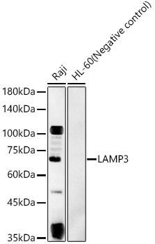
Western blot analysis of various lysates, using LAMP3 Rabbit pAb (CAB2895) at 1:3000 dilution. Secondary antibody: HRP-conjugated Goat anti-Rabbit IgG (H+L) (CABS014) at 1:10000 dilution. Lysates/proteins: 25μg per lane. Blocking buffer: 3% nonfat dry milk in TBST. Detection: ECL Basic Kit (AbGn00020). Exposure time: 180s.
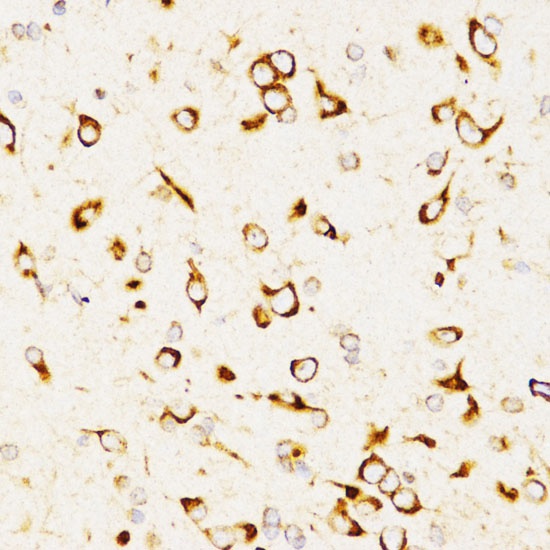
Immunohistochemistry analysis of paraffin-embedded Rat brain using LAMP3 Rabbit pAb (CAB2895) at dilution of 1:200 (40x lens). Microwave antigen retrieval performed with 0.01M PBS Buffer (pH 7.2) prior to IHC staining.