The Plastin L Monoclonal Antibody (CAB4664) is a high-quality antibody developed for reliable detection and analysis of target proteins. This antibody, produced in rabbit, exhibits high reactivity with human samples and is suitable for use in Western blot applications. By specifically binding to the L-Plastin protein, this antibody enables the detection and analysis of L-Plastin expression in various cell types, making it an essential component in studies related to cell motility, cancer metastasis, and immune cell function.L-Plastin, also known as LCP1, plays a crucial role in the regulation of actin dynamics and cell shape changes, making it an important factor in processes like cell migration and invasion.
This antibody is validated for use in WB, IHC-P, ELISA applications and has demonstrated reactivity against Human samples.
Product Name:
Plastin L Monoclonal Antibody
SKU:
CAB4664
Size:
20μL, 100μL
Reactivity:
Human
Clone Number:
ARC2689
Conjugate:
Unconjugated
Immunogen:
Synthetic peptide. This information is considered to be commercially sensitive.
Plastins are a family of actin-binding proteins that are conserved throughout eukaryote evolution and expressed in most tissues of higher eukaryotes. In humans, two ubiquitous plastin isoforms (L and T) have been identified. Plastin 1 (otherwise known as Fimbrin) is a third distinct plastin isoform which is specifically expressed at high levels in the small intestine. The L isoform is expressed only in hemopoietic cell lineages, while the T isoform has been found in all other normal cells of solid tissues that have replicative potential (fibroblasts, endothelial cells, epithelial cells, melanocytes, etc.). However, L-plastin has been found in many types of malignant human cells of non-hemopoietic origin suggesting that its expression is induced accompanying tumorigenesis in solid tissues.
Purification Method
Affinity purification
Gene ID
3936
Buffer Information
Store at -20℃. Avoid freeze / thaw cycles. Buffer: PBS containing 50% glycerol and 0.05% BSA, preserved with proclin300 or sodium azide, pH 7.3.
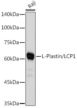
Western blot analysis of lysates from Raji cells, using L-Plastin/LCP1 Rabbit mAb (CAB4664) at 1:1000 dilution. Secondary antibody: HRP-conjugated Goat anti-Rabbit IgG (H+L) (CABS014) at 1:10000 dilution. Lysates/proteins: 25μg per lane. Blocking buffer: 3% nonfat dry milk in TBST. Detection: ECL Basic Kit (AbGn00020). Exposure time: 1s.
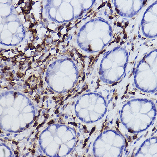
Immunohistochemistry analysis of paraffin-embedded Human colon using L-Plastin/LCP1 Rabbit mAb (CAB4664) at dilution of 1:100 (40x lens). High pressure antigen retrieval performed with 0.01M Citrate buffer (pH 6.0) prior to IHC staining.