The LCP2 Antibody (CAB2567) is a high-quality antibody developed for reliable detection and analysis of target proteins. This antibody, generated in rabbits, exhibits high specificity and sensitivity when detecting LCP2 in human samples, making it ideal for use in Western blot applications. LCP2, also known as lymphocyte cytosolic protein 2, plays a crucial role in T cell receptor signaling, leading to the activation of T cells and their response to antigens. Dysregulation of LCP2 has been implicated in various immune-related disorders, including autoimmune diseases and immune deficiencies.
This antibody is validated for use in WB, ELISA applications and has demonstrated reactivity against Human, Mouse samples.
Product Name:
LCP2 Antibody
SKU:
CAB2567
Size:
20μL, 100μL
Reactivity:
Human, Mouse
Conjugate:
Unconjugated
Immunogen:
Recombinant protein (or fragment).This information is considered to be commercially sensitive.
Recommended starting concentration is 1 μg/mL. Please optimize the concentration based on your specific assay requirements.
Synonyms:
IMD81, SLP76, SLP-76, LCP2
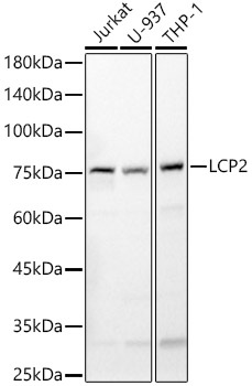
Positive Sample:
Jurkat, U-937, THP-1
Cellular Localization:
Cytoplasm.
Calculated MW:
60kDa
Observed MW:
76kDa
This gene encodes an adapter protein that acts as a substrate of the T cell antigen receptor (TCR)-activated protein tyrosine kinase pathway. The encoded protein associates with growth factor receptor bound protein 2, and is thought to play a role TCR-mediated intracellular signal transduction. A similar protein in mouse plays a role in normal T-cell development and activation. Mice lacking this gene show subcutaneous and intraperitoneal fetal hemorrhaging, dysfunctional platelets and impaired viability.
Purification Method
Affinity purification
Gene ID
3937
RRID
AB_2764455
Buffer Information
Store at -20℃. Avoid freeze / thaw cycles. Buffer: PBS containing 50% glycerol, preserved with proclin300 or sodium azide, pH 7.3.
Western blot analysis of various lysates, using LCP2 Rabbit pAb (CAB2567) at 1:500 dilution. Secondary antibody: HRP-conjugated Goat anti-Rabbit IgG (H+L) (CABS014) at 1:10000 dilution. Lysates/proteins: 25μg per lane. Blocking buffer: 3% nonfat dry milk in TBST. Detection: ECL Basic Kit (AbGn00020). Exposure time: 30s.