The SPN Antibody (CAB6412) is a high-quality antibody developed for reliable detection and analysis of target proteins. This polyclonal antibody, generated in rabbits, is highly specific to human leukosialin and is validated for use in a variety of research applications, including Western blotting.Leukosialin plays a crucial role in regulating immune cell interactions, including T cell activation and trafficking. Its expression has been linked to various immune-related diseases, making it an attractive target for investigation in immunology and cancer research.
This antibody is validated for use in WB, IHC-P, IF/ICC, ELISA applications and has demonstrated reactivity against Human, Mouse, Rat samples.
Product Name:
SPN Antibody
SKU:
CAB6412
Size:
20μL, 100μL
Reactivity:
Human, Mouse, Rat
Conjugate:
Unconjugated
Immunogen:
Recombinant protein (or fragment).This information is considered to be commercially sensitive.
Recommended starting concentration is 1 μg/mL. Please optimize the concentration based on your specific assay requirements.
Synonyms:
LSN, CD43, GALGP, GPL115, LEU-22, SPN
Positive Sample:
U-937
Cellular Localization:
Membrane, Single-Pass Type I Membrane Protein.
Calculated MW:
40kDa
Observed MW:
115-130kDa
This gene encodes a highly sialylated glycoprotein that functions in antigen-specific activation of T cells, and is found on the surface of thymocytes, T lymphocytes, monocytes, granulocytes, and some B lymphocytes. It contains a mucin-like extracellular domain, a transmembrane region and a carboxy-terminal intracellular region. The extracellular domain has a high proportion of serine and threonine residues, allowing extensive O-glycosylation, and has one potential N-glycosylation site, while the carboxy-terminal region has potential phosphorylation sites that may mediate transduction of activation signals. Different glycoforms of this protein have been described. In stimulated immune cells, proteolytic cleavage of the extracellular domain occurs in some cell types, releasing a soluble extracellular fragment. Defects in expression of this gene are associated with Wiskott-Aldrich syndrome.
Purification Method
Affinity purification
Gene ID
6693
RRID
AB_2767014
Buffer Information
Store at -20℃. Avoid freeze / thaw cycles. Buffer: PBS containing 50% glycerol, preserved with proclin300 or sodium azide, pH 7.3.
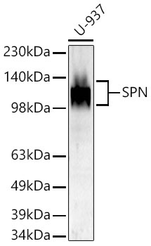
Western blot analysis of lysates from U-937 cells using SPN Rabbit pAb (CAB6412) at 1:1000 dilution incubated overnight at 4℃. Secondary antibody: HRP-conjugated Goat anti-Rabbit IgG (H+L) (CABS014) at 1:10000 dilution. Lysates/proteins: 25 μg per lane. Blocking buffer: 3% nonfat dry milk in TBST. Detection: ECL Basic Kit (AbGn00020). Exposure time: 90s.
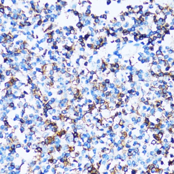
Immunohistochemistry analysis of paraffin-embedded Human tonsil using SPN Rabbit pAb (CAB6412) at dilution of 1:100 (40x lens). Microwave antigen retrieval performed with 0.01M PBS Buffer (pH 7.2) prior to IHC staining.
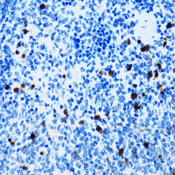
Immunohistochemistry analysis of paraffin-embedded Mouse spleen using SPN Rabbit pAb (CAB6412) at dilution of 1:100 (40x lens). Microwave antigen retrieval performed with 0.01M PBS Buffer (pH 7.2) prior to IHC staining.
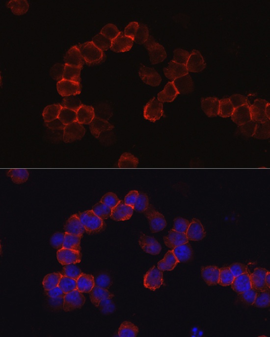
Immunofluorescence analysis of Jurkat cells using SPN Rabbit pAb (CAB6412) at dilution of 1:50 (40x lens). Secondary antibody: Cy3-conjugated Goat anti-Rabbit IgG (H+L) (CABS007) at 1:500 dilution. Blue: DAPI for nuclear staining.