The LIG3 Antibody (CAB13507) is a high-quality antibody developed for reliable detection and analysis of target proteins. This antibody, generated in rabbit hosts, shows high reactivity with human samples and is validated for use in techniques such as Western blotting. By specifically binding to Lig3 protein, this antibody enables accurate detection and analysis in various cell types, making it an invaluable asset for research in fields like genetics, molecular biology, and cancer studies.
This antibody is validated for use in WB, IHC-P, IF/ICC, ELISA applications and has demonstrated reactivity against Human, Mouse, Rat samples.
Product Name:
LIG3 Antibody
SKU:
CAB13507
Size:
20μL, 100μL
Reactivity:
Human, Mouse, Rat
Conjugate:
Unconjugated
Immunogen:
Recombinant protein (or fragment).This information is considered to be commercially sensitive.
Recommended starting concentration is 1 μg/mL. Please optimize the concentration based on your specific assay requirements.
Synonyms:
LIG2, MTDPS20, LIG3alpha, DNA Ligase III/LIG3
Positive Sample:
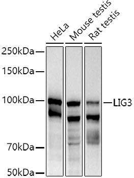
HeLa, Mouse testis, Rat testis
Cellular Localization:
Mitochondrion, Mitochondrion, Nucleus.
Calculated MW:
113kDa
Observed MW:
105kDa
This gene is a member of the DNA ligase family. Each member of this family encodes a protein that catalyzes the joining of DNA ends but they each have a distinct role in DNA metabolism. The protein encoded by this gene is involved in excision repair and is located in both the mitochondria and nucleus, with translation initiation from the upstream start codon allowing for transport to the mitochondria and translation initiation from a downstream start codon allowing for transport to the nucleus. Additionally, alternate transcriptional splice variants, encoding different isoforms, have been characterized.
Purification Method
Affinity purification
Gene ID
3980
RRID
AB_2760369
Buffer Information
Store at -20℃. Avoid freeze / thaw cycles. Buffer: PBS containing 50% glycerol, preserved with proclin300 or sodium azide, pH 7.3.
Western blot analysis of various lysates using (CAB13507) at 1:1000 dilution. Secondary antibody: HRP-conjugated Goat anti-Rabbit IgG (H+L) (CABS014) at 1:10000 dilution. Lysates/proteins: 25μg per lane. Blocking buffer: 3% nonfat dry milk in TBST. Detection: ECL Basic Kit (AbGn00020). Exposure time: 90s.
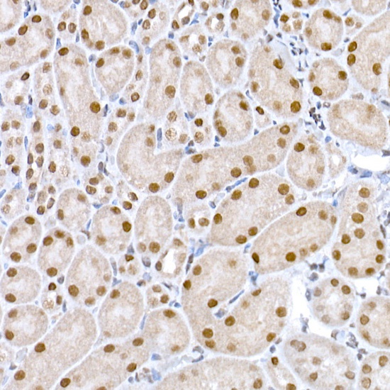
Immunohistochemistry analysis of paraffin-embedded Mouse kidney using DNA Ligase III/LIG3 Rabbit pAb (CAB13507) at dilution of 1:50 (40x lens). High pressure antigen retrieval performed with 0.01M Citrate buffer (pH 6.0) prior to IHC staining.
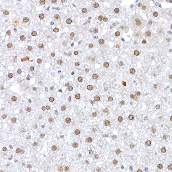
Immunohistochemistry analysis of paraffin-embedded Rat liver using DNA Ligase III/LIG3 Rabbit pAb (CAB13507) at dilution of 1:50 (40x lens). High pressure antigen retrieval performed with 0.01M Citrate buffer (pH 6.0) prior to IHC staining.
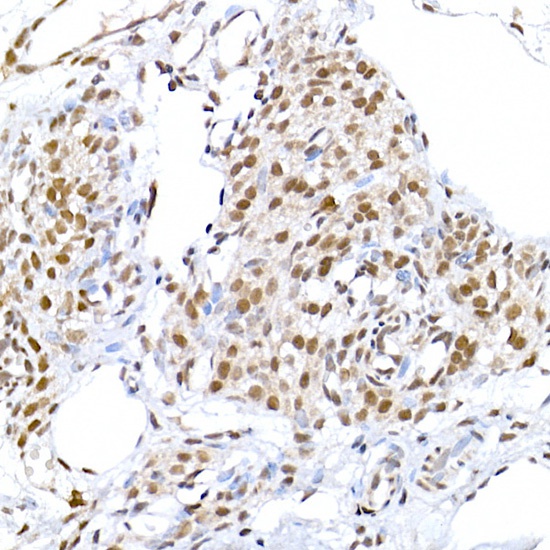
Immunohistochemistry analysis of paraffin-embedded Rat ovary using DNA Ligase III/LIG3 Rabbit pAb (CAB13507) at dilution of 1:50 (40x lens). High pressure antigen retrieval performed with 0.01M Citrate buffer (pH 6.0) prior to IHC staining.
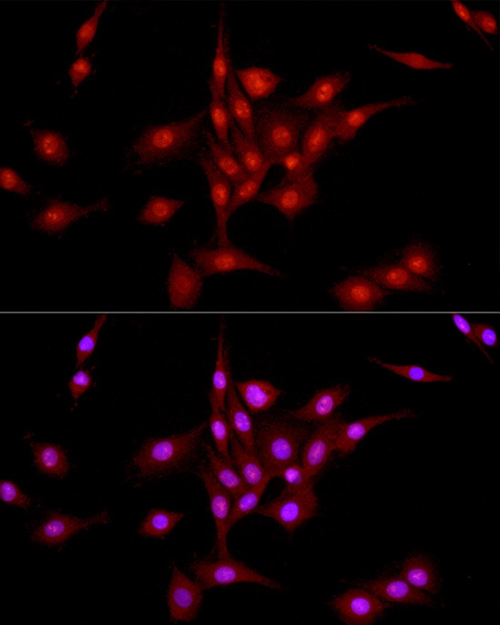
Immunofluorescence analysis of PC-12 cells using DNA Ligase III/LIG3 Rabbit pAb (CAB13507) at dilution of 1:50 (40x lens). Secondary antibody: Cy3-conjugated Goat anti-Rabbit IgG (H+L) (CABS007) at 1:500 dilution. Blue: DAPI for nuclear staining.