The LIMCH1 Antibody (CAB17649) is a high-quality antibody developed for reliable detection and analysis of target proteins. This antibody, produced in rabbits, exhibits high reactivity with human samples and has been validated for use in Western blot applications. By binding to the LIMCH1 protein, this antibody enables the detection and analysis of LIMCH1 in various cell types, making it an ideal choice for studies in cell biology and cancer research.
This antibody is validated for use in WB, ELISA applications and has demonstrated reactivity against Human samples.
Product Name:
LIMCH1 Antibody
SKU:
CAB17649
Size:
20μL, 100μL
Reactivity:
Human
Conjugate:
Unconjugated
Immunogen:
Recombinant protein (or fragment).This information is considered to be commercially sensitive.
Recommended starting concentration is 1 μg/mL. Please optimize the concentration based on your specific assay requirements.
Synonyms:
LMO7B, LIMCH1A, LIMCH1
Positive Sample:
LO2
Cellular Localization:
Cytoplasm.
Calculated MW:
122kDa
Observed MW:
140kDa
Enables myosin II head/neck binding activity. Involved in several processes, including cytoplasmic actin-based contraction involved in cell motility; positive regulation of stress fiber assembly; and regulation of focal adhesion assembly. Located in stress fiber. Colocalizes with myosin II complex.
Purification Method
Affinity purification
Gene ID
22998
RRID
AB_2770183
Buffer Information
Store at -20℃. Avoid freeze / thaw cycles. Buffer: PBS with 0.01% thimerosal,50% glycerol,pH7.3.
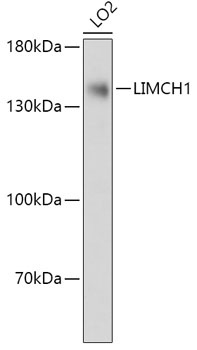
Western blot analysis of lysates from LO2 cells, using LIMCH1 Rabbit pAb (CAB17649) at 1:1000 dilution. Secondary antibody: HRP-conjugated Goat anti-Rabbit IgG (H+L) (CABS014) at 1:10000 dilution. Lysates/proteins: 25μg per lane. Blocking buffer: 3% nonfat dry milk in TBST. Detection: ECL Basic Kit (AbGn00020). Exposure time: 10s.