The LPP Monoclonal Antibody (CAB19226) is a high-quality antibody developed for reliable detection and analysis of target proteins. This antibody, raised in rabbits, demonstrates high reactivity with human samples and is validated for use in Western blot applications. By binding specifically to the LPP protein, researchers can accurately detect and analyze LPP expression in various cell types, making it an ideal choice for studies in cell biology and cancer research.LPP plays a critical role in cell adhesion and migration, making it a key player in processes such as wound healing, tissue regeneration, and cancer metastasis.
This antibody is validated for use in WB, IHC-P, IF/ICC, ELISA applications and has demonstrated reactivity against Human, Mouse samples.
Product Name:
LPP Monoclonal Antibody
SKU:
CAB19226
Size:
20μL, 100μL
Reactivity:
Human, Mouse
Clone Number:
ARC2381
Conjugate:
Unconjugated
Immunogen:
Synthetic peptide. This information is considered to be commercially sensitive.
Recommended starting concentration is 1 μg/mL. Please optimize the concentration based on your specific assay requirements.
Synonyms:
LPP, lipoma-preferred partner
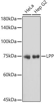
Positive Sample:
HeLa, Hep G2
Cellular Localization:
Cell Junction, Cell Membrane, Cytoplasm, Nucleus.
Calculated MW:
66kDa
Observed MW:
75kDa
This gene encodes a member of a subfamily of LIM domain proteins that are characterized by an N-terminal proline-rich region and three C-terminal LIM domains. The encoded protein localizes to the cell periphery in focal adhesions and may be involved in cell-cell adhesion and cell motility. This protein also shuttles through the nucleus and may function as a transcriptional co-activator. This gene is located at the junction of certain disease-related chromosomal translocations, which result in the expression of chimeric proteins that may promote tumor growth. Alternative splicing results in multiple transcript variants.
Purification Method
Affinity purification
Gene ID
4026
Buffer Information
Store at -20℃. Avoid freeze / thaw cycles. Buffer: PBS containing 50% glycerol and 0.05% BSA, preserved with proclin300 or sodium azide, pH 7.3.
Western blot analysis of various lysates using LPP Rabbit mAb (CAB19226) at 1:1000 dilution. Secondary antibody: HRP-conjugated Goat anti-Rabbit IgG (H+L) (CABS014) at 1:10000 dilution. Lysates/proteins: 25μg per lane. Blocking buffer: 3% nonfat dry milk in TBST. Detection: ECL Basic Kit (AbGn00020). Exposure time: 5s.
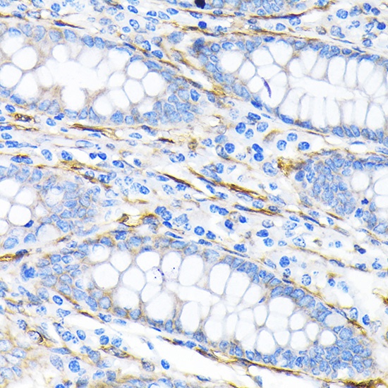
Immunohistochemistry analysis of paraffin-embedded Human liver cancer using LPP Rabbit mAb (CAB19226) at dilution of 1:100 (40x lens). Microwave antigen retrieval performed with 0.01M Tris/EDTA Buffer (pH 9.0) prior to IHC staining.