The LRG1 Antibody (CAB7850) is a high-quality antibody developed for reliable detection and analysis of target proteins. This antibody, generated from rabbit hosts, exhibits high reactivity towards human samples and has been validated for use in Western blot applications. By binding to the LRG1 protein, this antibody enables accurate detection and analysis in a variety of cell types, making it an essential tool for investigations in immunology and cancer research.
This antibody is validated for use in WB, ELISA, IF-P applications and has demonstrated reactivity against Human, Mouse, Rat samples.
Product Name:
LRG1 Antibody
SKU:
CAB7850
Size:
20μL, 100μL
Reactivity:
Human, Mouse, Rat
Conjugate:
Unconjugated
Immunogen:
Recombinant protein (or fragment).This information is considered to be commercially sensitive.
Recommended starting concentration is 1 μg/mL. Please optimize the concentration based on your specific assay requirements.
Synonyms:
LRG, HMFT1766, LRG1
Positive Sample:
Human plasma
Cellular Localization:
Secreted.
Calculated MW:
38kDa
Observed MW:
45kDa
The leucine-rich repeat (LRR) family of proteins, including LRG1, have been shown to be involved in protein-protein interaction, signal transduction, and cell adhesion and development. LRG1 is expressed during granulocyte differentiation (O'Donnell et al., 2002 [PubMed 12223515]).
Purification Method
Affinity purification
Gene ID
116844
RRID
AB_2770209
Buffer Information
Store at -20℃. Avoid freeze / thaw cycles. Buffer: PBS containing 50% glycerol, preserved with proclin300 or sodium azide, pH 7.3.
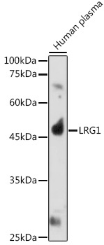
Western blot analysis of lysates from Human plasma, using LRG1 Rabbit pAb (CAB7850) at 1:1000 dilution. Secondary antibody: HRP-conjugated Goat anti-Rabbit IgG (H+L) (CABS014) at 1:10000 dilution. Lysates/proteins: 25μg per lane. Blocking buffer: 3% nonfat dry milk in TBST. Detection: ECL Basic Kit (AbGn00020). Exposure time: 180s.
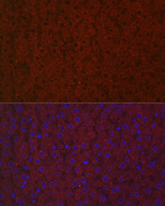
Immunofluorescence analysis of paraffin-embedded rat liver using LRG1 Rabbit pAb (CAB7850) at dilution of 1:100 (40x lens). Secondary antibody: Cy3-conjugated Goat anti-Rabbit IgG (H+L) (CABS007) at 1:500 dilution. Blue: DAPI for nuclear staining.
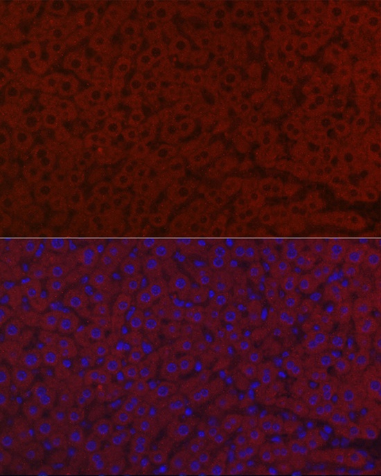
Immunofluorescence analysis of paraffin-embedded mouse liver using LRG1 Rabbit pAb (CAB7850) at dilution of 1:100 (40x lens). Secondary antibody: Cy3-conjugated Goat anti-Rabbit IgG (H+L) (CABS007) at 1:500 dilution. Blue: DAPI for nuclear staining.