The LRPAP1 Antibody (CAB13026) is a high-quality antibody developed for reliable detection and analysis of target proteins. This antibody, derived from rabbits, exhibits high reactivity with human samples and has been validated for use in Western blot applications.LRPAP1, also known as low density lipoprotein receptor-related protein-associated protein 1, plays a crucial role in the internalization and trafficking of LDL receptor-related proteins. Its involvement in cellular processes related to lipid metabolism and receptor signaling makes it a key target for studies in the fields of cardiovascular disease, lipid disorders, and neurodegenerative conditions.
This antibody is validated for use in WB, IHC-P, IF/ICC, ELISA applications and has demonstrated reactivity against Human, Mouse, Rat samples.
Product Name:
LRPAP1 Antibody
SKU:
CAB13026
Size:
20μL, 100μL
Reactivity:
Human, Mouse, Rat
Conjugate:
Unconjugated
Immunogen:
Recombinant protein (or fragment).This information is considered to be commercially sensitive.
This gene encodes a protein that interacts with the low density lipoprotein (LDL) receptor-related protein and facilitates its proper folding and localization by preventing the binding of ligands. Mutations in this gene have been identified in individuals with myopia 23. Alternative splicing results in multiple transcript variants.
Purification Method
Affinity purification
Gene ID
4043
RRID
AB_2759873
Buffer Information
Store at -20℃. Avoid freeze / thaw cycles. Buffer: PBS with 0.01% thimerosal,50% glycerol,pH7.3.
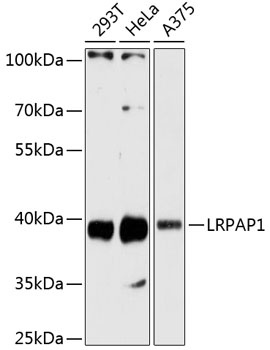
Western blot analysis of various lysates using LRPAP1 Rabbit pAb (CAB13026) at 1:3000 dilution. Secondary antibody: HRP-conjugated Goat anti-Rabbit IgG (H+L) (CABS014) at 1:10000 dilution. Lysates/proteins: 25μg per lane. Blocking buffer: 3% nonfat dry milk in TBST. Detection: ECL Basic Kit (AbGn00020). Exposure time: 60s.
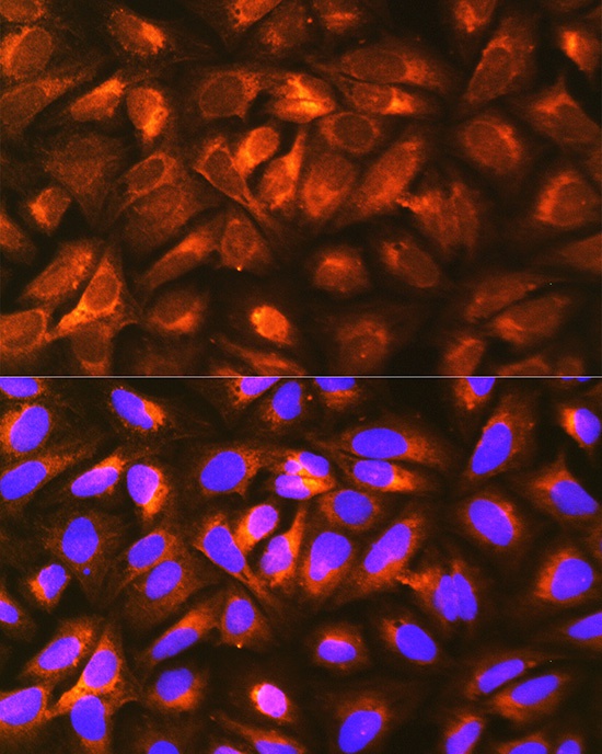
Immunofluorescence analysis of U-2 OS cells using LRPAP1 Rabbit pAb (CAB13026) at dilution of 1:100 (40x lens). Secondary antibody: Cy3-conjugated Goat anti-Rabbit IgG (H+L) (CABS007) at 1:500 dilution. Blue: DAPI for nuclear staining.