The Lumican (LUM) Antibody (CAB5352) is a high-quality antibody developed for reliable detection and analysis of target proteins. This polyclonal antibody, generated in rabbits, exhibits high reactivity with human samples and has been validated for use in Western blot applications. By specifically binding to the Lumican protein, researchers can accurately detect and analyze Lumican expression in various cell types, making it a versatile tool for studies in areas such as tissue development, wound healing, and cancer research.Lumican, a key component of the extracellular matrix, is involved in collagen fibril assembly and regulation of cell adhesion, migration, and proliferation.
This antibody is validated for use in WB, IF/ICC, ELISA applications and has demonstrated reactivity against Human, Mouse, Rat samples.
Product Name:
Lumican (LUM) Antibody
SKU:
CAB5352
Size:
20μL, 100μL
Reactivity:
Human, Mouse, Rat
Conjugate:
Unconjugated
Immunogen:
Recombinant protein (or fragment).This information is considered to be commercially sensitive.
This gene encodes a member of the small leucine-rich proteoglycan (SLRP) family that includes decorin, biglycan, fibromodulin, keratocan, epiphycan, and osteoglycin. In these bifunctional molecules, the protein moiety binds collagen fibrils and the highly charged hydrophilic glycosaminoglycans regulate interfibrillar spacings. Lumican is the major keratan sulfate proteoglycan of the cornea but is also distributed in interstitial collagenous matrices throughout the body. Lumican may regulate collagen fibril organization and circumferential growth, corneal transparency, and epithelial cell migration and tissue repair.
Purification Method
Affinity purification
Gene ID
4060
RRID
AB_2766162
Buffer Information
Store at -20℃. Avoid freeze / thaw cycles. Buffer: PBS with 0.09% Sodium azide,50% glycerol,pH7.3.
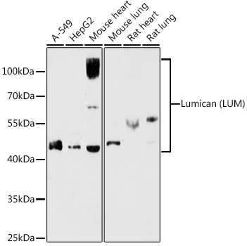
Western blot analysis of various lysates using Lumican (LUM) Rabbit pAb (CAB5352) at 1:1000 dilution. Secondary antibody: HRP-conjugated Goat anti-Rabbit IgG (H+L) (CABS014) at 1:10000 dilution. Lysates/proteins: 25μg per lane. Blocking buffer: 3% nonfat dry milk in TBST. Detection: ECL Basic Kit (AbGn00020). Exposure time: 180s.
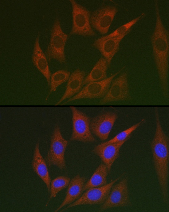
Immunofluorescence analysis of NIH/3T3 cells using Lumican (LUM) Rabbit pAb (CAB5352) at dilution of 1:100 (40x lens). Secondary antibody: Cy3-conjugated Goat anti-Rabbit IgG (H+L) (CABS007) at 1:500 dilution. Blue: DAPI for nuclear staining.


 ELISA Kit (MOEB1384)")
 ELISA Kit (RTEB1023)")
")
")