The LYPLA1 Antibody (CAB4419) is a high-quality antibody developed for reliable detection and analysis of target proteins. This antibody, generated in rabbits, exhibits high reactivity with human samples and has been validated for use in Western blot applications. By binding specifically to the LYPLA1 protein, researchers can accurately detect and analyze its expression in a variety of cell types, making it an excellent choice for studies in biochemistry, metabolism, and cell signaling.
This antibody is validated for use in WB, IHC-P, ELISA applications and has demonstrated reactivity against Human, Mouse samples.
Product Name:
LYPLA1 Antibody
SKU:
CAB4419
Size:
20μL, 100μL
Reactivity:
Human, Mouse
Conjugate:
Unconjugated
Immunogen:
Recombinant protein (or fragment).This information is considered to be commercially sensitive.
Recommended starting concentration is 1 μg/mL. Please optimize the concentration based on your specific assay requirements.
Synonyms:
APT1, LPL1, APT-1, LPL-I, hAPT1, LYPLA1
Positive Sample:
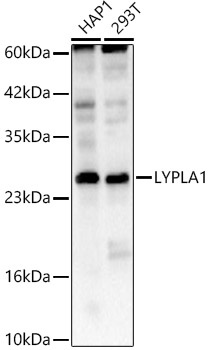
HAP1, 293T
Cellular Localization:
Cytoplasm.
Calculated MW:
25kDa
Observed MW:
25kDa
This gene encodes a member of the alpha/beta hydrolase superfamily. The encoded protein functions as a homodimer, exhibiting both depalmitoylating as well as lysophospholipase activity, and may be involved in Ras localization and signaling. Alternate splicing results in multiple transcript variants. Pseudogenes of this gene have been defined on chromosomes 4, 6, and 7.
Purification Method
Affinity purification
Gene ID
10434
RRID
AB_2765665
Buffer Information
Store at -20℃. Avoid freeze / thaw cycles. Buffer: PBS containing 50% glycerol, preserved with proclin300 or sodium azide, pH 7.3.
Western blot analysis of various lysates using LYPLA1 Rabbit pAb (CAB4419) at 1:1000 dilution. Secondary antibody: HRP-conjugated Goat anti-Rabbit IgG (H+L) (CABS014) at 1:10000 dilution. Lysates / proteins: 25 μg per lane. Blocking buffer: 3 % nonfat dry milk in TBST. Detection: ECL Basic Kit (AbGn00020). Exposure time: 90s.
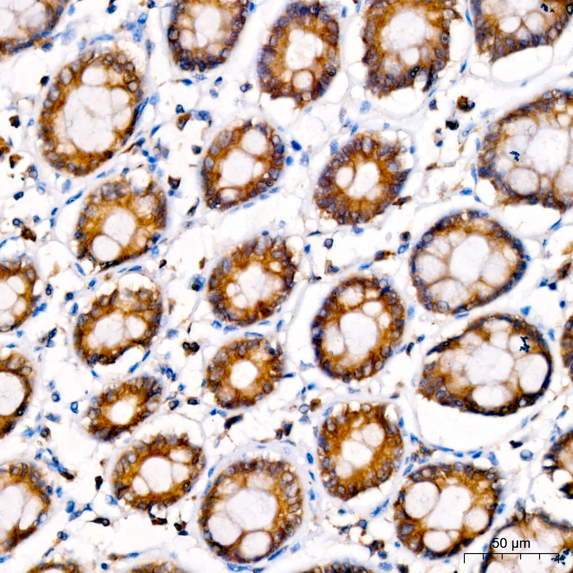
Immunohistochemistry analysis of paraffin-embedded Human colon tissue using LYPLA1 Rabbit pAb (CAB4419) at a dilution of 1:100 (40x lens). High pressure antigen retrieval was performed with 0.01 M citrate buffer (pH 6.0) prior to IHC staining.