The MAD2/MAD2L1 Monoclonal Antibody (CAB11469) is a high-quality antibody developed for reliable detection and analysis of target proteins. This antibody, produced in rabbits, demonstrates high specificity and sensitivity towards MAD2L1 in human samples, making it a reliable tool for Western blotting applications.MAD2L1, also known as mitotic arrest deficient 2 like 1, is a key player in the spindle assembly checkpoint, ensuring the accurate segregation of chromosomes during cell division. Dysregulation of MAD2L1 has been linked to various diseases, including cancer, making it a promising target for therapeutic interventions.
This antibody is validated for use in WB, ELISA applications and has demonstrated reactivity against Human samples.
Product Name:
MAD2/MAD2L1 Monoclonal Antibody
SKU:
CAB11469
Size:
20μL, 100μL
Reactivity:
Human
Clone Number:
ARC0603
Conjugate:
Unconjugated
Immunogen:
Synthetic peptide. This information is considered to be commercially sensitive.
Recommended starting concentration is 1 μg/mL. Please optimize the concentration based on your specific assay requirements.
Synonyms:
MAD2, HSMAD2, MAD2/MAD2L1
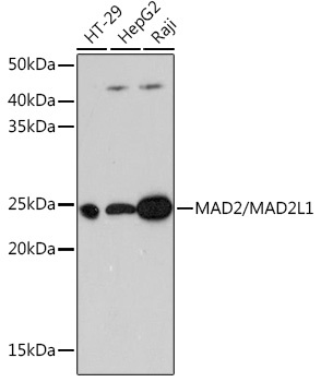
Positive Sample:
HT-29, HepG2, Raji
Cellular Localization:
Chromosome, Cytoplasm, Nucleus, Centromere, Cytoskeleton, Kinetochore, Spindle Pole.
Calculated MW:
24kDa
Observed MW:
24kDa
MAD2L1 is a component of the mitotic spindle assembly checkpoint that prevents the onset of anaphase until all chromosomes are properly aligned at the metaphase plate. MAD2L1 is related to the MAD2L2 gene located on chromosome 1. A MAD2 pseudogene has been mapped to chromosome 14.
Purification Method
Affinity purification
Gene ID
4085
RRID
AB_2861572
Buffer Information
Store at -20℃. Avoid freeze / thaw cycles. Buffer: PBS containing 50% glycerol and 0.05% BSA, preserved with proclin300 or sodium azide, pH 7.3.
Western blot analysis of various lysates using MAD2/MAD2/MAD2L1 Rabbit mAb (CAB11469) at 1:1000 dilution. Secondary antibody: HRP-conjugated Goat anti-Rabbit IgG (H+L) (CABS014) at 1:10000 dilution. Lysates/proteins: 25μg per lane. Blocking buffer: 3% nonfat dry milk in TBST. Detection: ECL Basic Kit (AbGn00020). Exposure time: 3min.


 (RPES1890)")
