The MAD2B/MAD2L2 Antibody (CAB12559) is a high-quality antibody developed for reliable detection and analysis of target proteins. This antibody, produced in rabbits, exhibits high reactivity with human samples and has been validated for use in Western blot applications. By binding specifically to the MAD2L2 protein, this antibody enables precise detection and analysis in various cell types, making it ideal for investigations in cell biology and cancer research.MAD2L2, also known as mitotic spindle assembly checkpoint protein, is essential for ensuring accurate chromosome segregation during cell division.
This antibody is validated for use in WB, IF/ICC, ELISA applications and has demonstrated reactivity against Human, Mouse, Rat samples.
Product Name:
MAD2B/MAD2L2 Antibody
SKU:
CAB12559
Size:
20μL, 100μL
Reactivity:
Human, Mouse, Rat
Conjugate:
Unconjugated
Immunogen:
Recombinant protein (or fragment).This information is considered to be commercially sensitive.
Recommended starting concentration is 1 μg/mL. Please optimize the concentration based on your specific assay requirements.
Synonyms:
REV7, FANCV, MAD2B, POLZ2, MAD2B/MAD2L2
Positive Sample:
SH-SY5Y, A375, K-562, SW480, HeLa, Mouse liver, Rat liver
Cellular Localization:
Cytoplasm, Nucleus, Cytoskeleton, Spindle.
Calculated MW:
24kDa
Observed MW:
24kDa
The protein encoded by this gene is a component of the mitotic spindle assembly checkpoint that prevents the onset of anaphase until all chromosomes are properly aligned at the metaphase plate. The encoded protein, which is similar to MAD2L1, is capable of interacting with ADAM9, ADAM15, REV1, and REV3 proteins.
Purification Method
Affinity purification
Gene ID
10459
RRID
AB_2759400
Buffer Information
Store at -20℃. Avoid freeze / thaw cycles. Buffer: PBS containing 50% glycerol, preserved with proclin300 or sodium azide, pH 7.3.
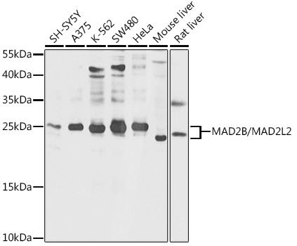
Western blot analysis of various lysates using MAD2B/MAD2L2 Rabbit pAb (CAB12559) at 1:1000 dilution. Secondary antibody: HRP-conjugated Goat anti-Rabbit IgG (H+L) (CABS014) at 1:10000 dilution. Lysates/proteins: 25μg per lane. Blocking buffer: 3% nonfat dry milk in TBST. Detection: ECL Basic Kit (AbGn00020). Exposure time: 5s.
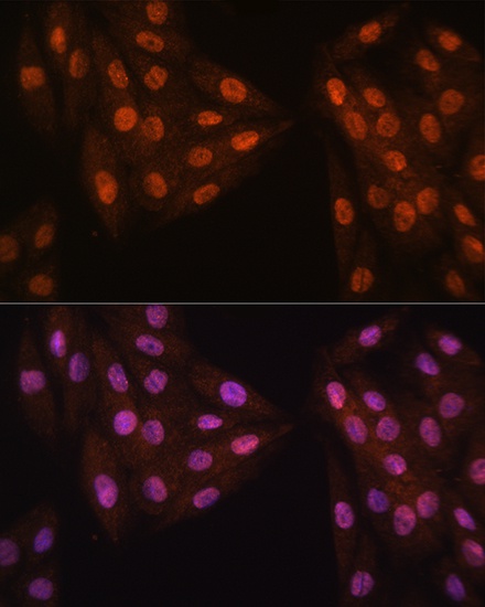
Immunofluorescence analysis of H9C2 cells using MAD2B/MAD2L2 Rabbit pAb (CAB12559) at dilution of 1:100. Blue: DAPI for nuclear staining.
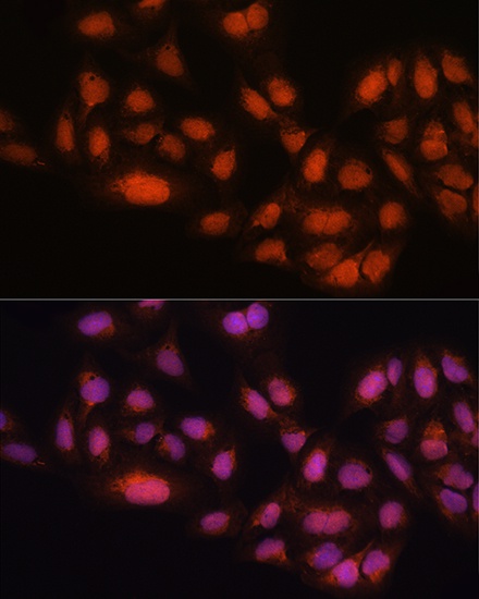
Immunofluorescence analysis of U2OS cells using MAD2B/MAD2L2 Rabbit pAb (CAB12559) at dilution of 1:100. Blue: DAPI for nuclear staining.