The MAGOHB Antibody (CAB16192) is a high-quality antibody developed for reliable detection and analysis of target proteins. The antibody, generated in rabbits, exhibits high specificity and sensitivity towards human samples, making it suitable for use in Western blot applications. By targeting the MAGOHB protein, this antibody allows for the detection and analysis of MAGOHB expression levels in various cell types, providing insights into its role in cellular processes.MAGOHB, a member of the MAGOH protein family, is known to be involved in mRNA export and localization, as well as in the regulation of alternative splicing events. Its functions are essential for maintaining proper gene expression and cellular functions, making it a key player in various biological processes.
This antibody is validated for use in WB, ELISA applications and has demonstrated reactivity against Human, Rat samples.
Product Name:
MAGOHB Antibody
SKU:
CAB16192
Size:
20μL, 100μL
Reactivity:
Human, Rat
Conjugate:
Unconjugated
Immunogen:
Recombinant protein (or fragment).This information is considered to be commercially sensitive.
Recommended starting concentration is 1 μg/mL. Please optimize the concentration based on your specific assay requirements.
Synonyms:
MGN2, mago, magoh, MAGOHB
Positive Sample:
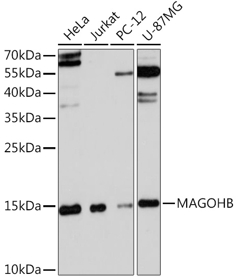
HeLa, Jurkat, PC-12, U-87MG
Cellular Localization:
Nucleus.
Calculated MW:
17kDa
Observed MW:
17kDa
Enables RNA binding activity. Involved in mRNA splicing, via spliceosome and nuclear-transcribed mRNA catabolic process, nonsense-mediated decay. Located in nucleus. Part of U2-type catalytic step 1 spliceosome; U2-type precatalytic spliceosome; and exon-exon junction complex.
Purification Method
Affinity purification
Gene ID
55110
RRID
AB_2763644
Buffer Information
Store at -20℃. Avoid freeze / thaw cycles. Buffer: PBS with 0.01% thimerosal,50% glycerol,pH7.3.
Western blot analysis of various lysates using MAGOHB Rabbit pAb (CAB16192) at 1:3000 dilution. Secondary antibody: HRP-conjugated Goat anti-Rabbit IgG (H+L) (CABS014) at 1:10000 dilution. Lysates/proteins: 25μg per lane. Blocking buffer: 3% nonfat dry milk in TBST. Detection: ECL Basic Kit (AbGn00020). Exposure time: 90s.