The MANF Antibody (CAB7005) is a high-quality antibody developed for reliable detection and analysis of target proteins. Raised in rabbits, this antibody is highly reactive with human samples and is validated for use in Western blot applications. It specifically binds to MANF, allowing for accurate detection and analysis in various cell types.MANF is a unique protein that has been shown to play a crucial role in protecting neurons from degeneration and promoting cell survival in conditions of stress.
This antibody is validated for use in WB, IHC-P, IF/ICC, IP, ELISA applications and has demonstrated reactivity against Human, Mouse, Rat samples.
Product Name:
MANF Antibody
SKU:
CAB7005
Size:
20μL, 100μL
Reactivity:
Human, Mouse, Rat
Conjugate:
Unconjugated
Immunogen:
Recombinant protein (or fragment).This information is considered to be commercially sensitive.
0.5μg-4μg antibody for 200μg-400μg extracts of whole cells
ELISA
Recommended starting concentration is 1 μg/mL. Please optimize the concentration based on your specific assay requirements.
Synonyms:
ARP, ARMET, ARMET/ARP/MANF
Positive Sample:
HT-29, HepG2, BxPC-3, Mouse testis, Rat liver, Rat testis, Rat pancreas
Cellular Localization:
Secreted.
Calculated MW:
21kDa
Observed MW:
16kDa
The protein encoded by this gene is localized in the endoplasmic reticulum (ER) and golgi, and is also secreted. Reducing expression of this gene increases susceptibility to ER stress-induced death and results in cell proliferation. Activity of this protein is important in promoting the survival of dopaminergic neurons. The presence of polymorphisms in the N-terminal arginine-rich region, including a specific mutation that changes an ATG start codon to AGG, have been reported in a variety of solid tumors; however, these polymorphisms were later shown to exist in normal tissues and are thus no longer thought to be tumor-related.
Purification Method
Affinity purification
Gene ID
7873
RRID
AB_2767561
Buffer Information
Store at -20℃. Avoid freeze / thaw cycles. Buffer: PBS containing 50% glycerol, preserved with proclin300 or sodium azide, pH 7.3.
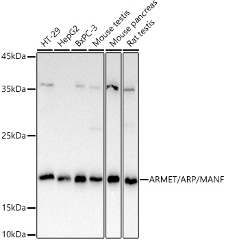
Western blot analysis of various lysates using ARMET/ARP/MANF Rabbit pAb (CAB7005) at 1:1000 dilution. Secondary antibody: HRP-conjugated Goat anti-Rabbit IgG (H+L) (CABS014) at 1:10000 dilution. Lysates/proteins: 25μg per lane. Blocking buffer: 3% nonfat dry milk in TBST. Detection: ECL Basic Kit (AbGn00020). Exposure time: 90s.
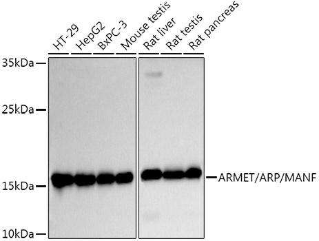
Western blot analysis of various lysates using ARMET/ARP/MANF Rabbit pAb (CAB7005) at 1:1000 dilution. Secondary antibody: HRP-conjugated Goat anti-Rabbit IgG (H+L) (CABS014) at 1:10000 dilution. Lysates/proteins: 25μg per lane. Blocking buffer: 3% nonfat dry milk in TBST. Detection: ECL Basic Kit (AbGn00020). Exposure time: 30s.
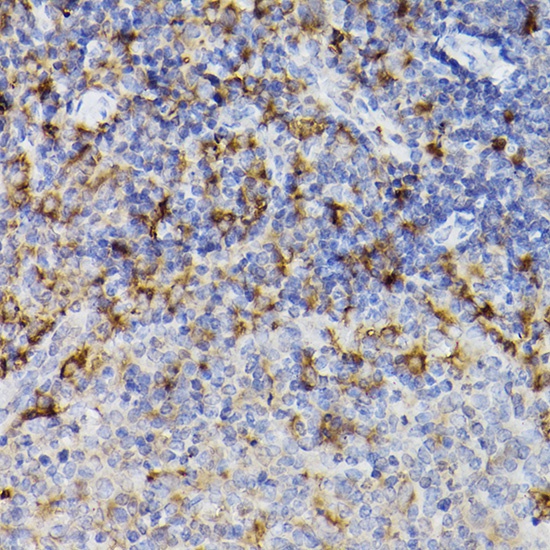
Immunohistochemistry analysis of paraffin-embedded Mouse spleen using ARMET/ARP/MANF Rabbit pAb (CAB7005) at dilution of 1:100 (40x lens). High pressure antigen retrieval performed with 0.01M Citrate buffer (pH 6.0) prior to IHC staining.
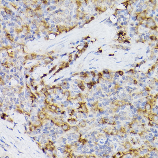
Immunohistochemistry analysis of paraffin-embedded Rat spleen using ARMET/ARP/MANF Rabbit pAb (CAB7005) at dilution of 1:100 (40x lens). High pressure antigen retrieval performed with 0.01M Citrate buffer (pH 6.0) prior to IHC staining.
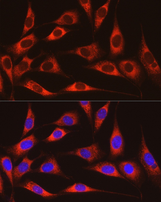
Immunofluorescence analysis of NIH/3T3 cells using ARMET/ARP/MANF Rabbit pAb (CAB7005) at dilution of 1:100 (40x lens). Secondary antibody: Cy3-conjugated Goat anti-Rabbit IgG (H+L) (CABS007) at 1:500 dilution. Blue: DAPI for nuclear staining.
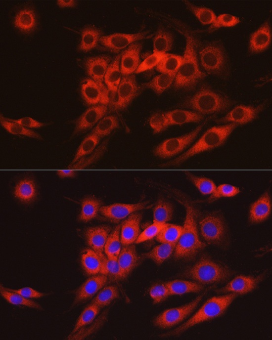
Immunofluorescence analysis of PC-12 cells using ARMET/ARP/MANF Rabbit pAb (CAB7005) at dilution of 1:100 (40x lens). Secondary antibody: Cy3-conjugated Goat anti-Rabbit IgG (H+L) (CABS007) at 1:500 dilution. Blue: DAPI for nuclear staining.
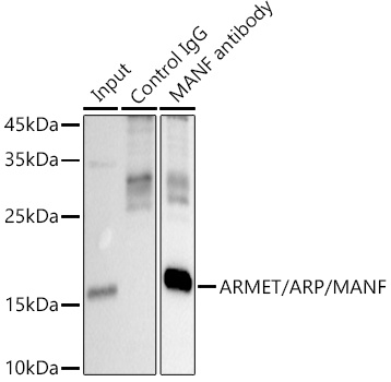
Immunoprecipitation analysis of 300 μg extracts of HepG2 cells using 3 μg ARMET/ARP/MANF antibody (CAB7005). Western blot was performed from the immunoprecipitate using ARMET/ARP/MANF antibody (CAB7005) at a dilution of 1:1000.