The MAP3K11 Antibody (CAB16242) is a high-quality antibody developed for reliable detection and analysis of target proteins. This antibody, raised in rabbits, is highly specific to human samples and has been validated for use in Western blot applications. By binding to the MAP3K11 protein, the antibody enables detection and analysis in various cell types, making it ideal for studies in immunology and cancer research.MAP3K11, also known as mixed-lineage kinase-3 (MLK3), is a key player in signaling cascades that regulate cell growth, survival, and differentiation.
This antibody is validated for use in WB, ELISA applications and has demonstrated reactivity against Mouse samples.
Product Name:
MAP3K11 Antibody
SKU:
CAB16242
Size:
20μL, 100μL
Reactivity:
Mouse
Immunogen:
Recombinant protein (or fragment).This information is considered to be commercially sensitive.
Recommended starting concentration is 1 μg/mL. Please optimize the concentration based on your specific assay requirements.
Synonyms:
MLK3, PTK1, SPRK, MLK-3, MEKK11, MAP3K11
Positive Sample:
Mouse thymus
Cellular Localization:
Centrosome, Cytosol.
Calculated MW:
93kDa
Observed MW:
93kDa
The protein encoded by this gene is a member of the serine/threonine kinase family. This kinase contains a SH3 domain and a leucine zipper-basic motif. This kinase preferentially activates MAPK8/JNK kinase, and functions as a positive regulator of JNK signaling pathway. This kinase can directly phosphorylate, and activates IkappaB kinase alpha and beta, and is found to be involved in the transcription activity of NF-kappaB mediated by Rho family GTPases and CDC42.
Purification Method
Affinity purification
Gene ID
4296
RRID
AB_2770265
Buffer Information
Store at -20℃. Avoid freeze / thaw cycles. Buffer: PBS with 0.01% thimerosal,50% glycerol,pH7.3.
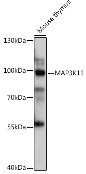
Western blot analysis of lysates from Mouse thymus, using MAP3K11 Rabbit pAb (CAB16242) at 1:1000 dilution. Secondary antibody: HRP-conjugated Goat anti-Rabbit IgG (H+L) (CABS014) at 1:10000 dilution. Lysates/proteins: 25μg per lane. Blocking buffer: 3% nonfat dry milk in TBST. Detection: ECL Basic Kit (AbGn00020). Exposure time: 1s.