The MAP4K2 Antibody (CAB17524) is a high-quality antibody developed for reliable detection and analysis of target proteins. This rabbit-derived antibody has been extensively validated for use in Western blot applications and is highly reactive with human samples, ensuring reliable and accurate results.MAP4K2, also known as mitogen-activated protein kinase kinase kinase kinase 2, is a critical regulator of various cellular processes, including cell growth, proliferation, and survival. Dysregulation of MAP4K2 activity has been implicated in several diseases, including cancer, inflammatory disorders, and autoimmune conditions.
This antibody is validated for use in WB, ELISA applications and has demonstrated reactivity against Human, Mouse, Rat samples.
Product Name:
MAP4K2 Antibody
SKU:
CAB17524
Size:
20μL, 100μL
Reactivity:
Human, Mouse, Rat
Conjugate:
Unconjugated
Immunogen:
Recombinant protein (or fragment).This information is considered to be commercially sensitive.
Recommended starting concentration is 1 μg/mL. Please optimize the concentration based on your specific assay requirements.
Synonyms:
GCK, BL44, RAB8IP, MAP4K2
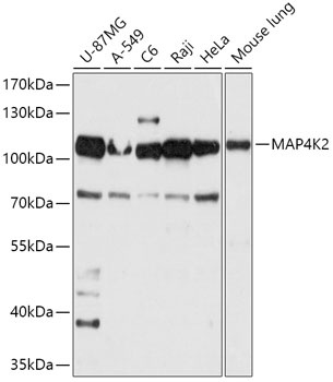
Positive Sample:
U-87MG, A-549, C6, Raji, HeLa, Mouse lung
Cellular Localization:
Basolateral Plasma Membrane, Cytoplasm.
Calculated MW:
92kDa
Observed MW:
110kDa
The protein encoded by this gene is a member of the serine/threonine protein kinase family. Although this kinase is found in many tissues, its expression in lymphoid follicles is restricted to the cells of germinal centre, where it may participate in B-cell differentiation. This kinase can be activated by TNF-alpha, and has been shown to specifically activate MAP kinases. This kinase is also found to interact with TNF receptor-associated factor 2 (TRAF2), which is involved in the activation of MAP3K1/MEKK1. Alternative splicing results in multiple transcript variants.
Purification Method
Affinity purification
Gene ID
5871
RRID
AB_2770272
Buffer Information
Store at -20℃. Avoid freeze / thaw cycles. Buffer: PBS with 0.01% thimerosal,50% glycerol,pH7.3.
Western blot analysis of various lysates using MAP4K2 Rabbit pAb (CAB17524) at 1:1000 dilution. Secondary antibody: HRP-conjugated Goat anti-Rabbit IgG (H+L) (CABS014) at 1:10000 dilution. Lysates/proteins: 25μg per lane. Blocking buffer: 3% nonfat dry milk in TBST. Detection: ECL Basic Kit (AbGn00020). Exposure time: 10s.