The ERK1/ERK2 Antibody (CAB16686) is a high-quality antibody developed for reliable detection and analysis of target proteins. This pathway plays a crucial role in various cellular processes, including cell growth, proliferation, differentiation, and apoptosis.Raised in rabbits, this antibody is highly specific and sensitive for detecting MAPK1 and MAPK3 in human samples. It has been validated for use in Western blot applications, allowing for precise and reliable detection of these important signaling molecules.Researchers in the fields of cell biology, molecular biology, and cancer research will benefit from the use of this antibody in their experiments.
This antibody is validated for use in WB, IHC-P, IF/ICC, ELISA applications and has demonstrated reactivity against Human, Mouse, Rat samples.
Product Name:
ERK1/ERK2 Antibody
SKU:
CAB16686
Size:
20μL, 100μL
Reactivity:
Human, Mouse, Rat
Conjugate:
Unconjugated
Immunogen:
Synthetic peptide. This information is considered to be commercially sensitive.
Sequence:
LNSK GYTK SIDI WSVG CILA EMLS NRPI FPGK HYLD QLNH ILGI LGSP SQED LNCI INLK ARNY LLSL PHKN KVPW NRLF PNAD SKAL DLLD KMLT FNPH K
Tested Applications:
WBIHC-PIF/ICCELISA
Recommended Dilution:
WB
1:500 - 1:5000
IF/ICC
1:50 - 1:200
IHC-P
1:50 - 1:200
ELISA
Recommended starting concentration is 1 μg/mL. Please optimize the concentration based on your specific assay requirements.
Synonyms:
MAPK1/MAPK3, ERK1 / ERK2
Positive Sample:
HeLa, 293T, C6, Mouse brain, Rat brain
Cellular Localization:
Caveola, Cytoplasm, Cytoskeleton, Cytosol, Early Endosome, Endoplasmic Reticulum Lumen, Extracellular Region, Focal Adhesion, Golgi Apparatus, Late Endosome, Microtubule Organizing Center, Mitochondrion, Mitotic Spindle, Nucleoplasm, Nucleus, Plasma Membrane.
Calculated MW:
36kDa/41kDa/38kDa/40kDa/43kDa
Observed MW:
42kDa/44kDa
MAP kinases, also known as extracellular signal-regulated kinases (ERKs), act as an integration point for multiple biochemical signals, and are involved in a wide variety of cellular processes such as proliferation, differentiation, transcription regulation and development. The activation of this kinase requires its phosphorylation by upstream kinases. Upon activation, this kinase translocates to the nucleus of the stimulated cells, where it phosphorylates nuclear targets.
Purification Method
Affinity purification
Gene ID
5594 5595
RRID
AB_2770274
Buffer Information
Store at -20℃. Avoid freeze / thaw cycles. Buffer: PBS containing 50% glycerol, preserved with proclin300 or sodium azide, pH 7.3.
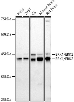
Western blot analysis of various lysates using ERK1 / ERK2 Rabbit pAb (CAB16686) at 1:1000 dilution. Secondary antibody: HRP-conjugated Goat anti-Rabbit IgG (H+L) (CABS014) at 1:10000 dilution. Lysates/proteins: 25μg per lane. Blocking buffer: 3% nonfat dry milk in TBST. Detection: ECL Basic Kit (AbGn00020). Exposure time: 90s.
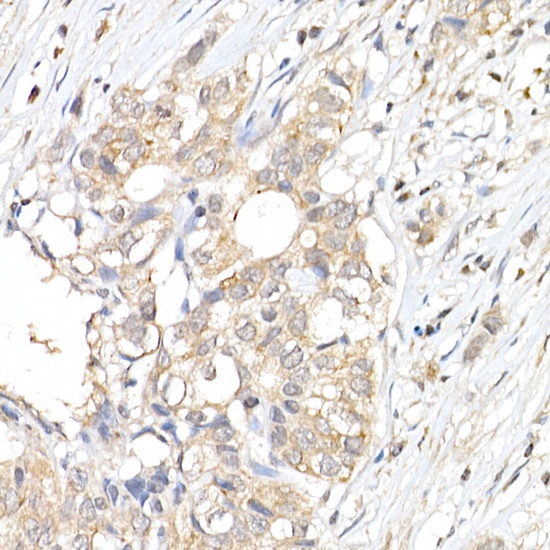
Immunohistochemistry analysis of paraffin-embedded Human breast cancer using ERK1 / ERK2 Rabbit pAb (CAB16686) at dilution of 1:50 (40x lens). High pressure antigen retrieval performed with 0.01M Citrate buffer (pH 6.0) prior to IHC staining.
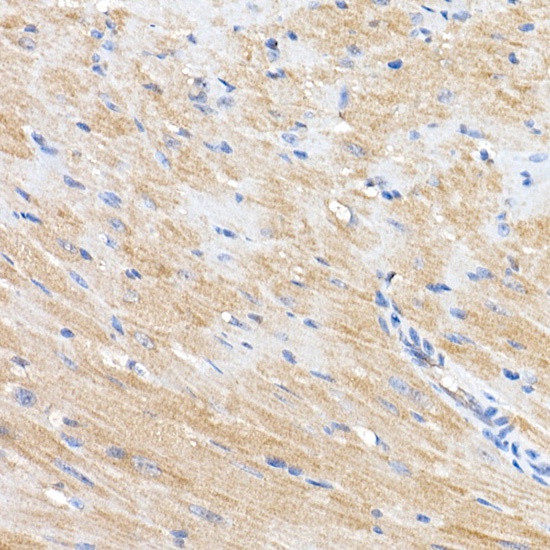
Immunohistochemistry analysis of paraffin-embedded Mouse heart using ERK1 / ERK2 Rabbit pAb (CAB16686) at dilution of 1:50 (40x lens). High pressure antigen retrieval performed with 0.01M Citrate buffer (pH 6.0) prior to IHC staining.
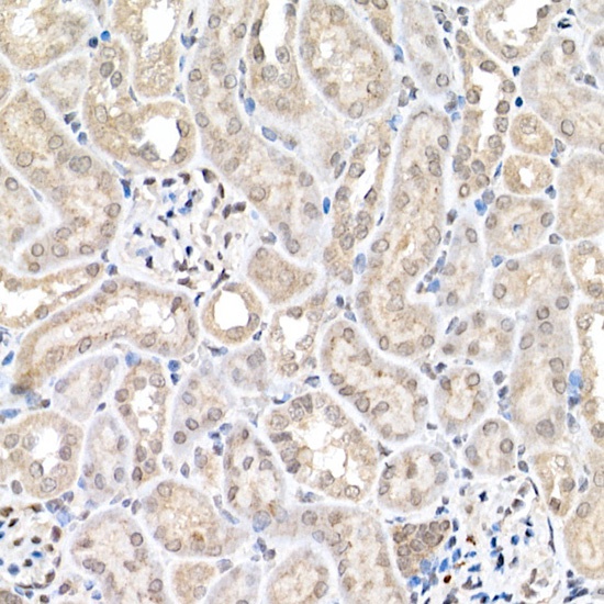
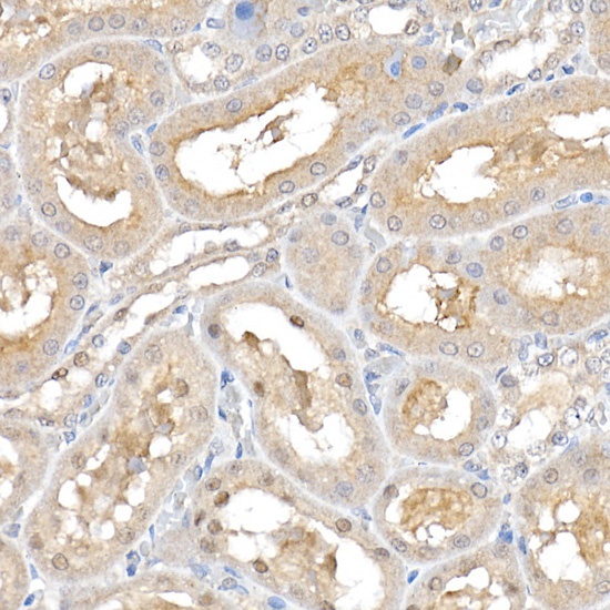
Immunohistochemistry analysis of paraffin-embedded Mouse kidney using ERK1 / ERK2 Rabbit pAb (CAB16686) at dilution of 1:50 (40x lens). High pressure antigen retrieval performed with 0.01M Citrate buffer (pH 6.0) prior to IHC staining.
Immunohistochemistry analysis of paraffin-embedded Rat kidney using ERK1 / ERK2 Rabbit pAb (CAB16686) at dilution of 1:50 (40x lens). High pressure antigen retrieval performed with 0.01M Citrate buffer (pH 6.0) prior to IHC staining.
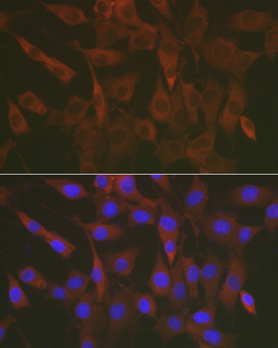
Immunofluorescence analysis of NIH/3T3 cells using ERK1 / ERK2 Rabbit pAb (CAB16686) at dilution of 1:50 (40x lens). Secondary antibody: Cy3-conjugated Goat anti-Rabbit IgG (H+L) (CABS007) at 1:500 dilution. Blue: DAPI for nuclear staining.