The MAPK11 Antibody (CAB7717) is a high-quality antibody developed for reliable detection and analysis of target proteins. This antibody, produced in rabbits, exhibits high reactivity with human samples and has been validated for use in Western blot applications. By specifically binding to the MAPK11 protein, this antibody enables precise detection and analysis in a variety of cell types, making it ideal for investigations in immunology, inflammation, and cancer research.MAPK11, also known as p38β, is a member of the MAPK signaling pathway that regulates a wide range of cellular processes, including cell proliferation, differentiation, and survival.
This antibody is validated for use in WB, IF/ICC, ELISA applications and has demonstrated reactivity against Human, Mouse, Rat samples.
Product Name:
MAPK11 Antibody
SKU:
CAB7717
Size:
20μL, 100μL
Reactivity:
Human, Mouse, Rat
Conjugate:
Unconjugated
Immunogen:
Synthetic peptide. This information is considered to be commercially sensitive.
This gene encodes a member of a family of protein kinases that are involved in the integration of biochemical signals for a wide variety of cellular processes, including cell proliferation, differentiation, transcriptional regulation, and development. The encoded protein can be activated by proinflammatory cytokines and environmental stresses through phosphorylation by mitogen activated protein kinase kinases (MKKs). Alternative splicing results in multiple transcript variants.
Purification Method
Affinity purification
Gene ID
5600
RRID
AB_2770278
Buffer Information
Store at -20℃. Avoid freeze / thaw cycles. Buffer: Buffer: PBS containing 50% glycerol, preserved with proclin300 or sodium azide, pH 7.3.
Western blot analysis of various lysates, using MAPK11 Rabbit pAb (CAB7717) at 1:800 dilution. Secondary antibody: HRP-conjugated Goat anti-Rabbit IgG (H+L) (CABS014) at 1:10000 dilution. Lysates/proteins: 25μg per lane. Blocking buffer: 3% nonfat dry milk in TBST. Detection: ECL Enhanced Kit (AbGn00021). Exposure time: 30s.
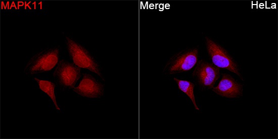
Immunofluorescence analysis of HeLa cells using MAPK11 Rabbit pAb (CAB7717) at a dilution of 1:50 (40x lens). Secondary antibody: Cy3-conjugated Goat anti-Rabbit IgG (H+L) (CABS007) at 1:500 dilution. Blue: DAPI for nuclear staining.
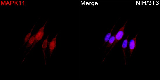
Immunofluorescence analysis of NIH/3T3 cells using MAPK11 Rabbit pAb(CAB7717) at a dilution of 1:50 (40x lens). Secondary antibody: Cy3-conjugated Goat anti-Rabbit IgG (H+L) (CABS007) at 1:500 dilution. Blue: DAPI for nuclear staining.
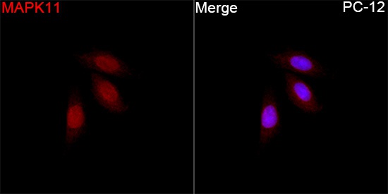
Immunofluorescence analysis of PC-12 cells using MAPK11 Rabbit pAb (CAB7717) at a dilution of 1:50 (40x lens). Secondary antibody: Cy3-conjugated Goat anti-Rabbit IgG (H+L) (CABS007) at 1:500 dilution. Blue: DAPI for nuclear staining.