The MAPRE1 Antibody (CAB2614) is a high-quality antibody developed for reliable detection and analysis of target proteins. This antibody specifically targets MAPRE1 and is raised in rabbits, ensuring high reactivity with human samples. Validated for use in Western blot applications, the antibody enables precise detection and analysis of MAPRE1 in various cell types.MAPRE1, also known as EB1, is a key regulator of microtubule dynamics, essential for processes such as cell migration, mitosis, and intracellular transport.
This antibody is validated for use in WB, IF/ICC, ELISA applications and has demonstrated reactivity against Human, Mouse, Rat samples.
Product Name:
MAPRE1 Antibody
SKU:
CAB2614
Size:
20μL, 100μL
Reactivity:
Human, Mouse, Rat
Conjugate:
Unconjugated
Immunogen:
Recombinant protein (or fragment).This information is considered to be commercially sensitive.
The protein encoded by this gene was first identified by its binding to the APC protein which is often mutated in familial and sporadic forms of colorectal cancer. This protein localizes to microtubules, especially the growing ends, in interphase cells. During mitosis, the protein is associated with the centrosomes and spindle microtubules. The protein also associates with components of the dynactin complex and the intermediate chain of cytoplasmic dynein. Because of these associations, it is thought that this protein is involved in the regulation of microtubule structures and chromosome stability. This gene is a member of the RP/EB family.
Purification Method
Affinity purification
Gene ID
22919
RRID
AB_2764493
Buffer Information
Store at -20℃. Avoid freeze / thaw cycles. Buffer: PBS with 0.01% thimerosal,50% glycerol,pH7.3.
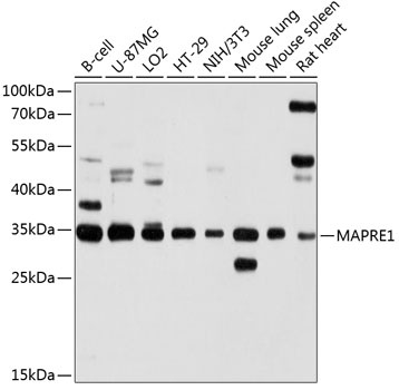
Western blot analysis of various lysates using MAPRE1 Rabbit pAb (CAB2614) at 1:1000 dilution. Secondary antibody: HRP-conjugated Goat anti-Rabbit IgG (H+L) (CABS014) at 1:10000 dilution. Lysates/proteins: 25μg per lane. Blocking buffer: 3% nonfat dry milk in TBST. Detection: ECL Basic Kit (AbGn00020). Exposure time: 10s.
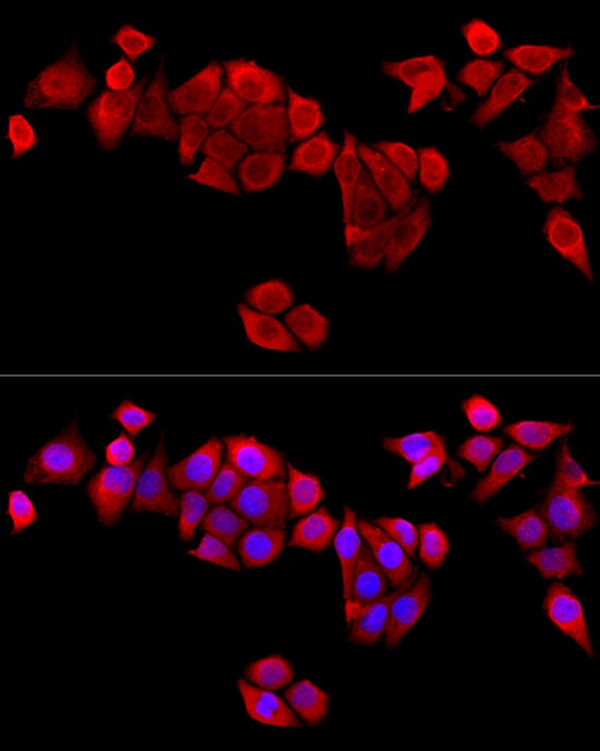
Immunofluorescence analysis of HeLa cells using MAPRE1 Rabbit pAb (CAB2614) at dilution of 1:100 (40x lens).Secondary antibody: Cy3-conjugated Goat anti-Rabbit IgG (H+L) (CABS007) at 1:500 dilution. Blue: DAPI for nuclear staining.