The MAT1A Antibody (CAB6650) is a high-quality antibody developed for reliable detection and analysis of target proteins. This antibody, derived from rabbit serum, exhibits high specificity and sensitivity in detecting MAT1A protein in human samples. Validated for use in various applications such as Western blot and immunohistochemistry, it enables researchers to accurately study the expression and localization of MAT1A in different cellular contexts.
This antibody is validated for use in WB, ELISA applications and has demonstrated reactivity against Human, Mouse, Rat samples.
Product Name:
MAT1A Antibody
SKU:
CAB6650
Size:
20μL, 100μL
Reactivity:
Human, Mouse, Rat
Conjugate:
Unconjugated
Immunogen:
Recombinant protein (or fragment).This information is considered to be commercially sensitive.
Recommended starting concentration is 1 μg/mL. Please optimize the concentration based on your specific assay requirements.
Synonyms:
MAT, SAMS, MATA1, SAMS1, MAT1A
Positive Sample:
Rat liver
Cellular Localization:
Cytosol.
Calculated MW:
44kDa
Observed MW:
44-55kDa
This gene catalyzes a two-step reaction that involves the transfer of the adenosyl moiety of ATP to methionine to form S-adenosylmethionine and tripolyphosphate, which is subsequently cleaved to PPi and Pi. S-adenosylmethionine is the source of methyl groups for most biological methylations. The encoded protein is found as a homotetramer (MAT I) or a homodimer (MAT III) whereas a third form, MAT II (gamma), is encoded by the MAT2A gene. Mutations in this gene are associated with methionine adenosyltransferase deficiency.
Purification Method
Affinity purification
Gene ID
4143
RRID
AB_2767238
Buffer Information
Store at -20℃. Avoid freeze / thaw cycles. Buffer: PBS containing 50% glycerol, preserved with proclin300 or sodium azide, pH 7.3.
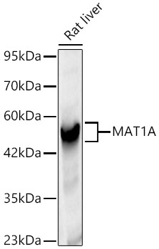
Western blot analysis of lysates from Rat liver, using MAT1A Rabbit pAb (CAB6650) at 1:1000 dilution. Secondary antibody: HRP-conjugated Goat anti-Rabbit IgG (H+L) (CABS014) at 1:10000 dilution. Lysates/proteins: 25μg per lane. Blocking buffer: 3% nonfat dry milk in TBST. Detection: ECL Basic Kit (AbGn00020). Exposure time: 5s.