The MC3R Antibody (CAB11609) is a high-quality antibody developed for reliable detection and analysis of target proteins. This antibody, generated in rabbits, is highly specific to human samples and has been validated for use in Western blot applications. By targeting the MC3R protein, this antibody allows for the accurate detection and analysis of MC3R expression in various cell types.The MC3R protein is a G protein-coupled receptor involved in the regulation of energy homeostasis and metabolism. It plays a crucial role in mediating the effects of melanocortin peptides, which are key regulators of appetite and energy expenditure.
This antibody is validated for use in WB, ELISA applications and has demonstrated reactivity against Mouse, Rat samples.
Product Name:
MC3R Antibody
SKU:
CAB11609
Size:
20μL, 100μL
Reactivity:
Mouse, Rat
Conjugate:
Unconjugated
Immunogen:
Recombinant protein (or fragment).This information is considered to be commercially sensitive.
Recommended starting concentration is 1 μg/mL. Please optimize the concentration based on your specific assay requirements.
Synonyms:
MC3, OB20, OQTL, BMIQ9, MC3-R, MC3R
Positive Sample:
Mouse brain, Mouse liver, Mouse kidney, Rat brain
Cellular Localization:
Cell Membrane, Multi-Pass Membrane Protein.
Calculated MW:
36kDa
Observed MW:
36kDa
This gene encodes a G-protein-coupled receptor for melanocyte-stimulating hormone and adrenocorticotropic hormone that is expressed in tissues other than the adrenal cortex and melanocytes. This gene maps to the same region as the locus for benign neonatal epilepsy. Mice deficient for this gene have increased fat mass despite decreased food intake, suggesting a role for this gene product in the regulation of energy homeostasis. Mutations in this gene are associated with a susceptibility to obesity in humans.
Purification Method
Affinity purification
Gene ID
4159
RRID
AB_2758633
Buffer Information
Store at -20℃. Avoid freeze / thaw cycles. Buffer: PBS with 0.01% thimerosal,50% glycerol,pH7.3.
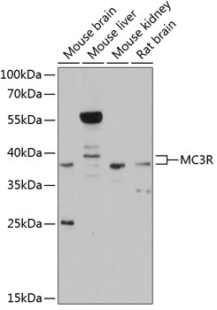
Western blot analysis of various lysates using MC3R Rabbit pAb (CAB11609) at 1:3000 dilution. Secondary antibody: HRP-conjugated Goat anti-Rabbit IgG (H+L) (CABS014) at 1:10000 dilution. Lysates/proteins: 25μg per lane. Blocking buffer: 3% nonfat dry milk in TBST. Detection: ECL Basic Kit (AbGn00020). Exposure time: 30s.