The CD146/MCAM Antibody (CAB13927) is a high-quality antibody developed for reliable detection and analysis of target proteins. This antibody, generated in rabbits, demonstrates high reactivity with human samples and is validated for use in Western blot applications. By binding specifically to the MCAM protein, researchers can accurately detect and analyze its expression in different cell types, making it an ideal choice for studies in cell biology, cancer research, and immunology.MCAM, also known as Melanoma Cell Adhesion Molecule, plays a key role in cell adhesion and migration processes, making it a potential target for therapeutic interventions in cancer metastasis.
This antibody is validated for use in WB, IHC-P, IF/ICC, ELISA applications and has demonstrated reactivity against Human, Mouse, Rat samples.
Product Name:
CD146/MCAM Antibody
SKU:
CAB13927
Size:
20μL, 100μL
Reactivity:
Human, Mouse, Rat
Conjugate:
Unconjugated
Immunogen:
Recombinant protein (or fragment).This information is considered to be commercially sensitive.
Recommended starting concentration is 1 μg/mL. Please optimize the concentration based on your specific assay requirements.
Synonyms:
CD146, MUC18, HEMCAM, METCAM, MelCAM, CD146/MCAM
Positive Sample:
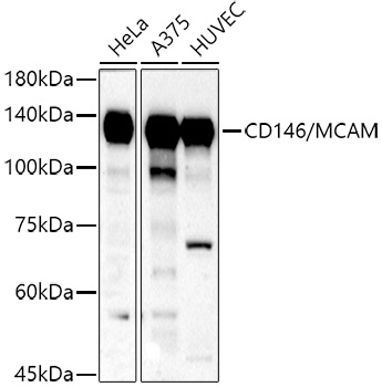
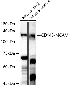
HeLa, A375, HUVEC, Mouse lung, Mouse uterus
Cellular Localization:
Membrane, Single-Pass Type I Membrane Protein.
Calculated MW:
72kDa
Observed MW:
120kDa
Involved in glomerular filtration and vascular wound healing. Acts upstream of or within angiogenesis. Located in external side of plasma membrane. Biomarker of chronic obstructive pulmonary disease and uveal melanoma.
Purification Method
Affinity purification
Gene ID
4162
RRID
AB_2760779
Buffer Information
Store at -20℃. Avoid freeze / thaw cycles. Buffer: PBS containing 50% glycerol, preserved with proclin300 or sodium azide, pH 7.3.
Western blot analysis of various lysates using CD146/MCAM Rabbit pAb (CAB13927) at 1:500 dilution. Secondary antibody: HRP-conjugated Goat anti-Rabbit IgG (H+L) (CABS014) at 1:10000 dilution. Lysates/proteins: 25μg per lane. Blocking buffer: 3% nonfat dry milk in TBST. Detection: ECL Basic Kit (AbGn00020). Exposure time: 180s.
Western blot analysis of various lysates using CD146/MCAM Rabbit pAb (CAB13927) at 1:500 dilution. Secondary antibody: HRP-conjugated Goat anti-Rabbit IgG (H+L) (CABS014) at 1:10000 dilution. Lysates/proteins: 25μg per lane. Blocking buffer: 3% nonfat dry milk in TBST. Detection: ECL Enhanced Kit (AbGn00021). Exposure time: 180s.
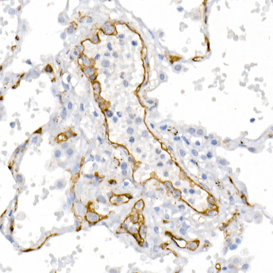
Immunohistochemistry analysis of paraffin-embedded Human lung cancer using CD146/MCAM Rabbit pAb (CAB13927) at dilution of 1:20 (40x lens). High pressure antigen retrieval performed with 0.01M Citrate buffer (pH 6.0) prior to IHC staining.
Immunohistochemistry analysis of paraffin-embedded Human lung using CD146/MCAM Rabbit pAb (CAB13927) at dilution of 1:20 (40x lens). High pressure antigen retrieval performed with 0.01M Citrate buffer (pH 6.0) prior to IHC staining.
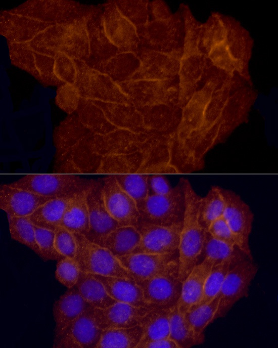
Immunofluorescence analysis of HeLa cells using CD146/MCAM Rabbit pAb (CAB13927) at dilution of 1:50 (40x lens). Secondary antibody: Cy3-conjugated Goat anti-Rabbit IgG (H+L) (CABS007) at 1:500 dilution. Blue: DAPI for nuclear staining.


")
")