The MCCC1 Antibody (CAB10020) is a high-quality antibody developed for reliable detection and analysis of target proteins. This antibody, produced in rabbits, exhibits high reactivity with human samples and has been validated for use in Western blot applications. By specifically binding to the MCCC1 protein, this antibody allows for precise detection and analysis in a variety of cell types, making it an essential component for studies in metabolism and related disorders.MCCC1 is responsible for the conversion of 3-methylcrotonyl-CoA to 3-methylglutaconyl-CoA, a crucial step in the breakdown of certain amino acids and fatty acids for energy production.
This antibody is validated for use in WB, IHC-P, ELISA applications and has demonstrated reactivity against Human, Mouse, Rat samples.
Product Name:
MCCC1 Antibody
SKU:
CAB10020
Size:
20μL, 100μL
Reactivity:
Human, Mouse, Rat
Conjugate:
Unconjugated
Immunogen:
Recombinant protein (or fragment).This information is considered to be commercially sensitive.
Sequence:
VSSQ ETQG GPLA PMTG TIEK VFVK AGDK VKAG DSLM VMIA MKME HTIK SPKD GTVK KVFY REGA QANR HTPL VEFE EEES DKRE SE
Tested Applications:
WBIHC-PELISA
Recommended Dilution:
WB
1:1000 - 1:5000
IHC-P
1:50 - 1:100
ELISA
Recommended starting concentration is 1 μg/mL. Please optimize the concentration based on your specific assay requirements.
Synonyms:
MCCA, MCC-B, MCCCalpha, MCCC1
Positive Sample:
T-47D
Cellular Localization:
Mitochondrion Matrix.
Calculated MW:
80kDa
Observed MW:
726aa/80kDa
This gene encodes the large subunit of 3-methylcrotonyl-CoA carboxylase. This enzyme functions as a heterodimer and catalyzes the carboxylation of 3-methylcrotonyl-CoA to form 3-methylglutaconyl-CoA. Mutations in this gene are associated with 3-Methylcrotonylglycinuria, an autosomal recessive disorder of leucine catabolism.
Purification Method
Affinity purification
Gene ID
56922
RRID
AB_2757540
Buffer Information
Store at -20℃. Avoid freeze / thaw cycles. Buffer: PBS containing 50% glycerol, preserved with proclin300 or sodium azide, pH 7.3.
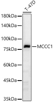
Western blot analysis of lysates from T-47D cells, using MCCC1 Rabbit pAb (CAB10020) at 1:2000 dilution. Secondary antibody: HRP-conjugated Goat anti-Rabbit IgG (H+L) (CABS014) at 1:10000 dilution. Lysates/proteins: 25μg per lane. Blocking buffer: 3% nonfat dry milk in TBST. Detection: ECL Basic Kit (AbGn00020). Exposure time: 60s.
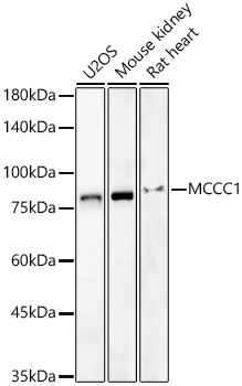
Western blot analysis of various lysates, using MCCC1 Rabbit pAb (CAB10020) at 1:2000 dilution. Secondary antibody: HRP-conjugated Goat anti-Rabbit IgG (H+L) (CABS014) at 1:10000 dilution. Lysates/proteins: 25μg per lane. Blocking buffer: 3% nonfat dry milk in TBST. Detection: ECL Basic Kit (AbGn00020). Exposure time: 60s.
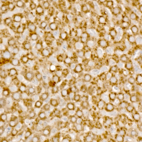
Immunohistochemistry analysis of paraffin-embedded Rat liver using MCCC1 Rabbit pAb (CAB10020) at dilution of 1:100 (40x lens). High pressure antigen retrieval performed with 0.01M Citrate buffer (pH 6.0) prior to IHC staining.