The MCL1 Antibody (CAB0250) is a high-quality antibody developed for reliable detection and analysis of target proteins. This antibody, generated in rabbits, has high reactivity with human samples and is validated for use in Western blot applications. By binding specifically to the MCL1 protein, this antibody allows for accurate detection and analysis in various cell types, making it ideal for investigations in cancer research and cell biology.MCL1, a member of the Bcl-2 family of proteins, plays a crucial role in controlling cell survival and resistance to apoptosis. Its dysregulation is often associated with cancer development and progression, making it an important target for therapeutic interventions.
This antibody is validated for use in WB, IHC-P, IF/ICC, ELISA applications and has demonstrated reactivity against Human, Mouse samples.
Product Name:
MCL1 Antibody
SKU:
CAB0250
Size:
20μL, 100μL
Reactivity:
Human, Mouse
Conjugate:
Unconjugated
Immunogen:
Synthetic peptide. This information is considered to be commercially sensitive.
This gene encodes an anti-apoptotic protein, which is a member of the Bcl-2 family. Alternative splicing results in multiple transcript variants. The longest gene product (isoform 1) enhances cell survival by inhibiting apoptosis while the alternatively spliced shorter gene products (isoform 2 and isoform 3) promote apoptosis and are death-inducing.
Purification Method
Affinity purification
Gene ID
4170
RRID
AB_2757063
Buffer Information
Store at -20℃. Avoid freeze / thaw cycles. Buffer: PBS containing 50% glycerol, preserved with proclin300 or sodium azide, pH 7.3.
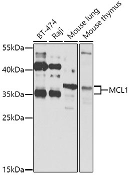
Western blot analysis of various lysates using MCL1 Rabbit pAb (CAB0250) at 1:1000 dilution. Secondary antibody: HRP-conjugated Goat anti-Rabbit IgG (H+L) (CABS014) at 1:10000 dilution. Lysates/proteins: 25μg per lane. Blocking buffer: 3% nonfat dry milk in TBST. Detection: ECL Basic Kit (AbGn00020). Exposure time: 30s.
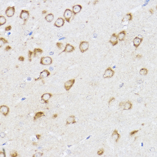
Immunohistochemistry analysis of paraffin-embedded Mouse brain using MCL1 Rabbit pAb (CAB0250) at dilution of 1:100 (40x lens). High pressure antigen retrieval performed with 0.01M Citrate buffer (pH 6.0) prior to IHC staining.