The MCU Monoclonal Antibody (CAB22525) is a high-quality antibody developed for reliable detection and analysis of target proteins. This antibody, developed through monoclonal antibody technology, exhibits high specificity and sensitivity for detecting MCU in various biological samples.The MCU protein is a key player in mitochondrial calcium uptake, a process essential for cellular bioenergetics and signaling. Dysregulation of MCU has been implicated in numerous diseases, including neurodegenerative disorders, cardiovascular diseases, and cancer. By targeting MCU with this monoclonal antibody, researchers can gain valuable insights into the mechanisms underlying these pathologies.
This antibody is validated for use in WB, ELISA applications and has demonstrated reactivity against Human, Mouse, Rat samples.
Product Name:
MCU Monoclonal Antibody
SKU:
CAB22525
Size:
20μL, 100μL
Reactivity:
Human, Mouse, Rat
Clone Number:
ARC57879
Conjugate:
Unconjugated
Immunogen:
Recombinant protein (or fragment).This information is considered to be commercially sensitive.
Recommended starting concentration is 1 μg/mL. Please optimize the concentration based on your specific assay requirements.
Synonyms:
HsMCU, C10orf42, CCDC109A, CU
Positive Sample:
HeLa, 293T, NIH/3T3, Mouse spleen, Rat skeletal muscle
Cellular Localization:
Mitochondrial Inner Membrane, Mitochondrion.
Calculated MW:
40kDa
Observed MW:
35kDa
Enables calcium channel activity; identical protein binding activity; and uniporter activity. Involved in several processes, including positive regulation of mitochondrial calcium ion concentration; positive regulation of mitochondrial fission; and positive regulation of neutrophil chemotaxis. Acts upstream of or within calcium import into the mitochondrion. Located in mitochondrial inner membrane. Is integral component of mitochondrial inner membrane. Part of uniplex complex.
Purification Method
Affinity purification
Gene ID
90550
Buffer Information
Store at -20℃. Avoid freeze / thaw cycles. Buffer: PBS with 0.09% Sodium azide,0.05% BSA,50% glycerol,pH7.3.
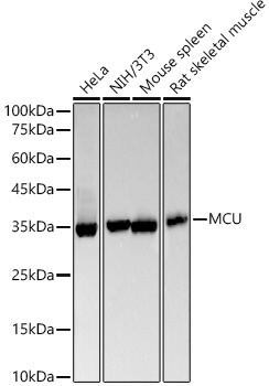
Western blot analysis of various lysates, using [KO Validated] MCU Rabbit mAb (CAB22525) at1:20000 dilution. Secondary antibody: HRP-conjugated Goat anti-Rabbit IgG (H+L) (CABS014) at 1:10000 dilution. Lysates/proteins: 25μg per lane. Blocking buffer: 3% nonfat dry milk in TBST. Detection: ECL Basic Kit (AbGn00020). Exposure time: 90s.
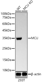
Western blot analysis of lysates from wild type(WT) and MCU knockout (KO) 293T(KO) cells, using [KO Validated] MCU Rabbit mAb (CAB22525) at1:20000 dilution. Secondary antibody: HRP-conjugated Goat anti-Rabbit IgG (H+L) (CABS014) at 1:10000 dilution. Lysates/proteins: 25μg per lane. Blocking buffer: 3% nonfat dry milk in TBST. Detection: ECL Basic Kit (AbGn00020). Exposure time: 90s.
 at1:20000 dilution. Secondary antibody: HRP Goat Anti-Rabbit IgG (H+L) at 1:10000 dilution. Lysates/proteins: 25μg per lane. Blocking buffer: 3% nonfat dry milk in TBST.")
 at1:20000 dilution. Secondary antibody: HRP Goat Anti-Rabbit IgG (H+L) at 1:10000 dilution. Lysates/proteins: 25μg per lane. Blocking buffer: 3% nonfat dry milk in TBST.")

