The MDK Antibody (CAB0251) is a high-quality antibody developed for reliable detection and analysis of target proteins. This antibody, generated in rabbits, exhibits high specificity and sensitivity for detecting Midkine in human samples.Midkine is a growth factor that plays a crucial role in promoting cell proliferation and survival, making it a key player in cancer development and progression. By targeting Midkine with this antibody, researchers can investigate its role in tumorigenesis and potentially identify new therapeutic targets for cancer treatment.This antibody is validated for use in Western blot applications, allowing for the detection and analysis of Midkine expression in different cell types and tissues.
This antibody is validated for use in WB, IF/ICC, ELISA applications and has demonstrated reactivity against Human, Mouse, Rat samples.
Product Name:
MDK Antibody
SKU:
CAB0251
Size:
20μL, 100μL
Reactivity:
Human, Mouse, Rat
Conjugate:
Unconjugated
Immunogen:
Recombinant protein (or fragment).This information is considered to be commercially sensitive.
Recommended starting concentration is 1 μg/mL. Please optimize the concentration based on your specific assay requirements.
Synonyms:
MK, ARAP, NEGF2, MDK
Positive Sample:
SH-SY5Y
Cellular Localization:
Secreted.
Calculated MW:
16kDa
Observed MW:
16kDa
This gene encodes a member of a small family of secreted growth factors that binds heparin and responds to retinoic acid. The encoded protein promotes cell growth, migration, and angiogenesis, in particular during tumorigenesis. This gene has been targeted as a therapeutic for a variety of different disorders. Alternatively spliced transcript variants encoding multiple isoforms have been observed.
Purification Method
Affinity purification
Gene ID
4192
RRID
AB_2757064
Buffer Information
Store at -20℃. Avoid freeze / thaw cycles. Buffer: PBS containing 50% glycerol, preserved with proclin300 or sodium azide, pH 7.3.
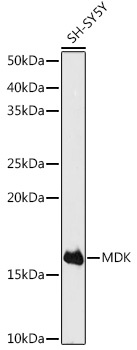
Western blot analysis of lysates from SH-SY5Y cells, using MDK Rabbit pAb (CAB0251) at 1:500 dilution. Secondary antibody: HRP-conjugated Goat anti-Rabbit IgG (H+L) (CABS014) at 1:10000 dilution. Lysates/proteins: 25μg per lane. Blocking buffer: 3% nonfat dry milk in TBST. Detection: ECL Enhanced Kit (AbGn00021). Exposure time: 180s.
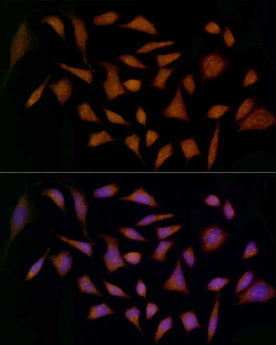
Immunofluorescence analysis of HeLa cells using MDK Rabbit pAb (CAB0251) at dilution of 1:20 (40x lens). Secondary antibody: Cy3-conjugated Goat anti-Rabbit IgG (H+L) (CABS007) at 1:500 dilution. Blue: DAPI for nuclear staining.