The MERTK Antibody (CAB5443) is a high-quality antibody developed for reliable detection and analysis of target proteins. This antibody, produced in rabbits, exhibits high reactivity with human samples and is validated for use in Western blot applications. By binding specifically to the MERTK protein, this antibody enables precise detection and analysis in a variety of cell types, making it an essential resource for studies in immunology and cancer research.MERTK is a key player in the regulation of immune responses and the clearance of apoptotic cells, making it a promising target for investigation in diseases such as cancer, autoimmune disorders, and chronic inflammatory conditions.
This antibody is validated for use in WB, IHC-P, IF/ICC, ELISA applications and has demonstrated reactivity against Human, Mouse, Rat samples.
Product Name:
MERTK Antibody
SKU:
CAB5443
Size:
20μL, 100μL
Reactivity:
Human, Mouse, Rat
Conjugate:
Unconjugated
Immunogen:
Recombinant protein (or fragment).This information is considered to be commercially sensitive.
Recommended starting concentration is 1 μg/mL. Please optimize the concentration based on your specific assay requirements.
Synonyms:
MER, RP38, c-Eyk, c-mer, Tyro12, MERTK
Positive Sample:
HepG2, Mouse spleen
Cellular Localization:
Membrane, Single-Pass Type I Membrane Protein.
Calculated MW:
110kDa
Observed MW:
150-200kDa
This gene is a member of the MER/AXL/TYRO3 receptor kinase family and encodes a transmembrane protein with two fibronectin type-III domains, two Ig-like C2-type (immunoglobulin-like) domains, and one tyrosine kinase domain. Mutations in this gene have been associated with disruption of the retinal pigment epithelium (RPE) phagocytosis pathway and onset of autosomal recessive retinitis pigmentosa (RP).
Purification Method
Affinity purification
Gene ID
10461
RRID
AB_2766245
Buffer Information
Store at -20℃. Avoid freeze / thaw cycles. Buffer: PBS containing 50% glycerol, preserved with proclin300 or sodium azide, pH 7.3.
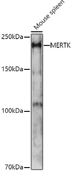
Western blot analysis of lysates from Mouse spleen, using MERTK Rabbit pAb (CAB5443) at 1:1000 dilution. Secondary antibody: HRP-conjugated Goat anti-Rabbit IgG (H+L) (CABS014) at 1:10000 dilution. Lysates/proteins: 25μg per lane. Blocking buffer: 3% nonfat dry milk in TBST. Detection: ECL Enhanced Kit (AbGn00021). Exposure time: 180s.
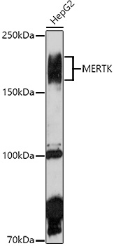
Western blot analysis of lysates from HepG2 cells, using MERTK Rabbit pAb (CAB5443) at 1:1000 dilution. Secondary antibody: HRP-conjugated Goat anti-Rabbit IgG (H+L) (CABS014) at 1:10000 dilution. Lysates/proteins: 25μg per lane. Blocking buffer: 3% nonfat dry milk in TBST. Detection: ECL Enhanced Kit (AbGn00021). Exposure time: 180s.
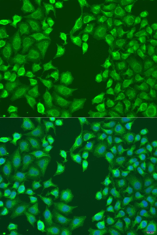
Immunofluorescence analysis of U2OS cells using MERTK Rabbit pAb (CAB5443) at dilution of 1:100 (40x lens). Secondary antibody: Cy3-conjugated Goat anti-Rabbit IgG (H+L) (CABS007) at 1:500 dilution. Blue: DAPI for nuclear staining.