The METTL7A Antibody (CAB8201) is a high-quality antibody developed for reliable detection and analysis of target proteins. This antibody, produced in rabbits, is highly specific to human samples and has been validated for use in various applications, including Western blotting.Mettl7a is an essential component of the RNA methyltransferase complex, playing a critical role in the regulation of mRNA stability and translation efficiency. Its involvement in these processes makes it a promising target for studying post-transcriptional gene regulation and the molecular mechanisms underlying various diseases, including cancer and neurodegenerative disorders.
This antibody is validated for use in WB, IHC-P, IF/ICC, ELISA applications and has demonstrated reactivity against Human, Mouse, Rat samples.
Product Name:
METTL7A Antibody
SKU:
CAB8201
Size:
20μL, 100μL
Reactivity:
Human, Mouse, Rat
Conjugate:
Unconjugated
Immunogen:
Recombinant protein (or fragment).This information is considered to be commercially sensitive.
Recommended starting concentration is 1 μg/mL. Please optimize the concentration based on your specific assay requirements.
Synonyms:
AAMB, AAM-B, METTL7A
Positive Sample:
22Rv1
Cellular Localization:
Endoplasmic Reticulum, Lipid Droplet, Membrane.
Calculated MW:
28kDa
Observed MW:
28kDa
Predicted to enable methyltransferase activity. Predicted to be involved in methylation. Located in lipid droplet.
Purification Method
Affinity purification
Gene ID
25840
RRID
AB_2770347
Buffer Information
Store at -20℃. Avoid freeze / thaw cycles. Buffer: PBS containing 50% glycerol, preserved with proclin300 or sodium azide, pH 7.3.
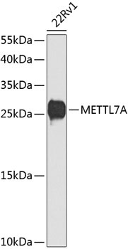
Western blot analysis of lysates from 22Rv1 cells, using METTL7A Rabbit pAb (CAB8201) at 1:1000 dilution. Secondary antibody: HRP-conjugated Goat anti-Rabbit IgG (H+L) (CABS014) at 1:10000 dilution. Lysates/proteins: 25μg per lane. Blocking buffer: 3% nonfat dry milk in TBST. Detection: ECL Basic Kit (AbGn00020). Exposure time: 90s.
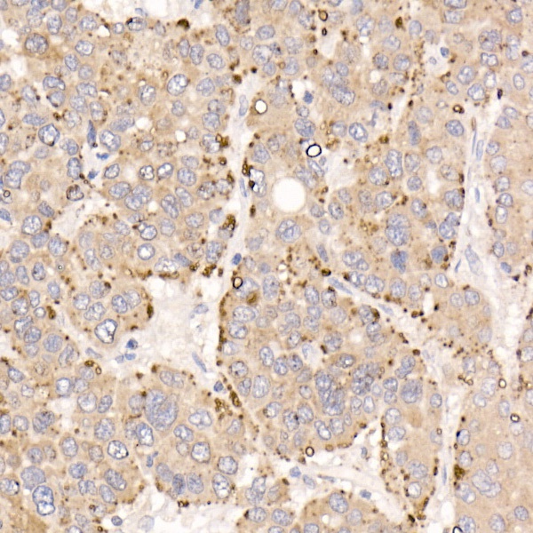
Immunohistochemistry analysis of paraffin-embedded Human liver cancer using METTL7A Rabbit pAb (CAB8201) at dilution of 1:20 (40x lens). High pressure antigen retrieval performed with 0.01M Citrate buffer (pH 6.0) prior to IHC staining.
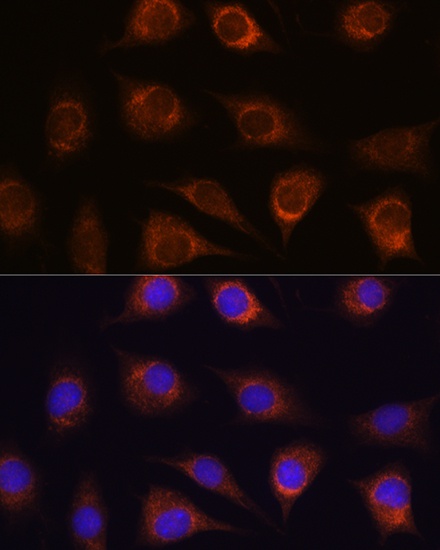
Immunofluorescence analysis of L929 cells using METTL7A Rabbit pAb (CAB8201) at dilution of 1:100. Secondary antibody: Cy3-conjugated Goat anti-Rabbit IgG (H+L) (CABS007) at 1:500 dilution. Blue: DAPI for nuclear staining.
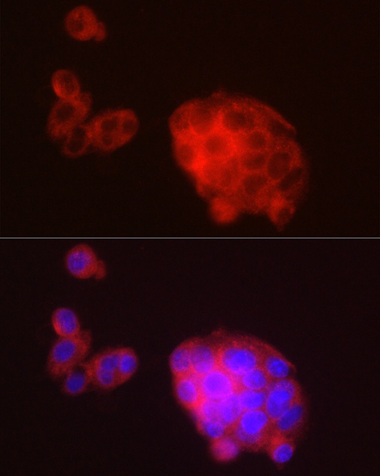
Immunofluorescence analysis of HepG2 cells using METTL7A Rabbit pAb (CAB8201) at dilution of 1:100 (40x lens). Secondary antibody: Cy3-conjugated Goat anti-Rabbit IgG (H+L) (CABS007) at 1:500 dilution. Blue: DAPI for nuclear staining.