The MGAT3 Antibody (CAB8134) is a high-quality antibody developed for reliable detection and analysis of target proteins. This antibody, produced through rabbit immunization, exhibits high reactivity with human samples and is validated for use in Western blot applications. By binding specifically to the MGAT3 protein, researchers can accurately detect and analyze its expression in various cell types, making it an essential resource for studies in glycobiology and cancer research.MGAT3 plays a crucial role in the maturation and branching of N-glycans, impacting various biological processes such as cell adhesion, signaling, and immune response modulation.
This antibody is validated for use in WB, ELISA applications and has demonstrated reactivity against Human, Mouse, Rat samples.
Product Name:
MGAT3 Antibody
SKU:
CAB8134
Size:
20μL, 100μL
Reactivity:
Human, Mouse, Rat
Conjugate:
Unconjugated
Immunogen:
Recombinant protein (or fragment).This information is considered to be commercially sensitive.
Recommended starting concentration is 1 μg/mL. Please optimize the concentration based on your specific assay requirements.
Synonyms:
GNT3, GNT-III, MGAT3
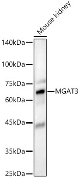
Positive Sample:
Mouse kidney
Cellular Localization:
Golgi Apparatus Membrane, Single-Pass Type Ii Membrane Protein.
Calculated MW:
61kDa
Observed MW:
61kDa
There are believed to be over 100 different glycosyltransferases involved in the synthesis of protein-bound and lipid-bound oligosaccharides. The enzyme encoded by this gene transfers a GlcNAc residue to the beta-linked mannose of the trimannosyl core of N-linked oligosaccharides and produces a bisecting GlcNAc. Multiple alternatively spliced variants, encoding the same protein, have been identified.
Purification Method
Affinity purification
Gene ID
4248
RRID
AB_2770356
Buffer Information
Store at -20℃. Avoid freeze / thaw cycles. Buffer: PBS containing 50% glycerol, preserved with proclin300 or sodium azide, pH 7.3.
Western blot analysis of lysates from Mouse kidney, using MGAT3 Rabbit pAb (CAB8134) at 1:1000 dilution. Secondary antibody: HRP-conjugated Goat anti-Rabbit IgG (H+L) (CABS014) at 1:10000 dilution. Lysates/proteins: 25μg per lane. Blocking buffer: 3% nonfat dry milk in TBST. Detection: ECL Basic Kit (AbGn00020). Exposure time: 180s.