The MGP Antibody (CAB5439) is a high-quality antibody developed for reliable detection and analysis of target proteins. Raised in rabbits, this antibody demonstrates high reactivity with human samples and is validated for use in various applications including Western blotting.Matrix Gla Protein is known for its role in preventing pathological calcification of soft tissues, particularly in blood vessels. Dysregulation of MGP expression has been linked to conditions such as arterial calcification, atherosclerosis, and cardiovascular disease. By targeting MGP, researchers can investigate its function in maintaining vascular health and explore potential therapeutic interventions for cardiovascular disorders.
This antibody is validated for use in WB, IHC-P, ELISA, IF-P applications and has demonstrated reactivity against Human, Mouse, Rat samples.
Product Name:
MGP Antibody
SKU:
CAB5439
Size:
20μL, 100μL
Reactivity:
Human, Mouse, Rat
Conjugate:
Unconjugated
Immunogen:
Recombinant protein (or fragment).This information is considered to be commercially sensitive.
Recommended starting concentration is 1 μg/mL. Please optimize the concentration based on your specific assay requirements.
Synonyms:
NTI, GIG36, MGLAP, MGP
Positive Sample:
SH-SY5Y
Cellular Localization:
Secreted.
Calculated MW:
12kDa
Observed MW:
12kDa
This gene encodes a member of the osteocalcin/matrix Gla family of proteins. The encoded vitamin K-dependent protein is secreted by chondrocytes and vascular smooth muscle cells, and functions as a physiological inhibitor of ectopic tissue calcification. Carboxylation status of the encoded protein is associated with calcification of the vasculature in human patients with cardiovascular disease and calcification of the synovial membranes in osteoarthritis patients. Mutations in this gene cause Keutel syndrome in human patients, which is characterized by abnormal cartilage calcification, peripheral pulmonary stenosis and facial hypoplasia.
Purification Method
Affinity purification
Gene ID
4256
RRID
AB_2766241
Buffer Information
Store at -20℃. Avoid freeze / thaw cycles. Buffer: PBS containing 50% glycerol, preserved with proclin300 or sodium azide, pH 7.3.
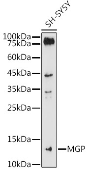
Western blot analysis of lysates from SH-SY5Y cells, using MGP Rabbit pAb (CAB5439) at 1:1000 dilution. Secondary antibody: HRP-conjugated Goat anti-Rabbit IgG (H+L) (CABS014) at 1:10000 dilution. Lysates/proteins: 25μg per lane. Blocking buffer: 3% nonfat dry milk in TBST. Detection: ECL Basic Kit (AbGn00020). Exposure time: 180s.
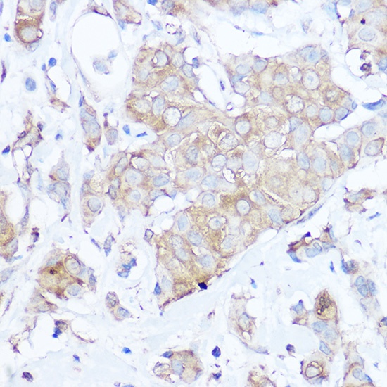
Immunohistochemistry analysis of paraffin-embedded Human breast cancer using MGP Rabbit pAb (CAB5439) at dilution of 1:100 (40x lens). High pressure antigen retrieval performed with 0.01M Citrate buffer (pH 6.0) prior to IHC staining.
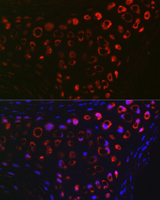
Immunofluorescence analysis of paraffin-embedded Rat cartilage using MGP Rabbit pAb (CAB5439) at dilution of 1:100 (40x lens). Secondary antibody: Cy3-conjugated Goat anti-Rabbit IgG (H+L) (CABS007) at 1:500 dilution. Blue: DAPI for nuclear staining.
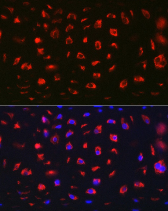
Immunofluorescence analysis of paraffin-embedded Mouse cartilage using MGP Rabbit pAb (CAB5439) at dilution of 1:100 (40x lens). Secondary antibody: Cy3-conjugated Goat anti-Rabbit IgG (H+L) (CABS007) at 1:500 dilution. Blue: DAPI for nuclear staining.