The MIER1 Antibody (CAB18480) is a high-quality antibody developed for reliable detection and analysis of target proteins. This antibody, raised in rabbits, is highly specific and reacts strongly with human samples, making it an excellent choice for Western blot applications. It binds specifically to the MIER1 protein, allowing for accurate detection and analysis in a variety of cell types.MIER1, also known as mesoderm induction early response 1, is involved in various cellular processes, including development, differentiation, and cell growth.
This antibody is validated for use in WB, IHC-P, ELISA applications and has demonstrated reactivity against Human, Mouse samples.
Product Name:
MIER1 Antibody
SKU:
CAB18480
Size:
20μL, 100μL
Reactivity:
Human, Mouse
Immunogen:
Recombinant protein (or fragment).This information is considered to be commercially sensitive.
Recommended starting concentration is 1 μg/mL. Please optimize the concentration based on your specific assay requirements.
Synonyms:
ER1, MI-ER1, MIER1
Positive Sample:
HeLa, A-549, Mouse brain
Cellular Localization:
Cytoplasm, Nucleoplasm, Nucleus.
Calculated MW:
58kDa
Observed MW:
58kDa
This gene encodes a protein that was first identified in Xenopus laevis by its role in a mesoderm induction early response (MIER). The encoded protein functions as a transcriptional regulator. Alternatively spliced transcript variants encode multiple isoforms, some of which lack a C-terminal nuclear localization signal.
Purification Method
Affinity purification
Gene ID
57708
RRID
AB_2862247
Buffer Information
Store at -20℃. Avoid freeze / thaw cycles. Buffer: PBS with 0.01% thimerosal,50% glycerol,pH7.3.
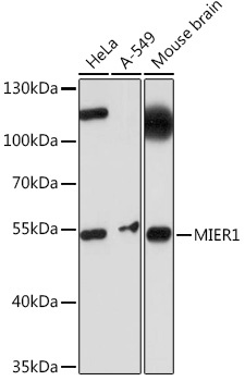
Western blot analysis of various lysates using MIER1 Rabbit pAb (CAB18480) at 1:1000 dilution. Secondary antibody: HRP-conjugated Goat anti-Rabbit IgG (H+L) (CABS014) at 1:10000 dilution. Lysates/proteins: 25μg per lane. Blocking buffer: 3% nonfat dry milk in TBST. Detection: ECL Basic Kit (AbGn00020). Exposure time: 10s.
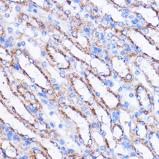
Immunohistochemistry analysis of paraffin-embedded Mouse kidney using MIER1 Rabbit pAb (CAB18480) at dilution of 1:100 (40x lens). Microwave antigen retrieval performed with 0.01M PBS Buffer (pH 7.2) prior to IHC staining.