The MIF Antibody (CAB1391) is a high-quality antibody developed for reliable detection and analysis of target proteins. This antibody, raised in rabbits, is highly specific and reactive with human samples, making it an ideal choice for experiments involving MIF detection and analysis.MIF is a pro-inflammatory cytokine that plays a key role in various biological processes, including immune responses, inflammation, and cell proliferation. By targeting the MIF protein, researchers can gain valuable insights into the mechanisms underlying these processes and potentially identify new therapeutic targets for diseases like cancer, autoimmune disorders, and inflammatory conditions.
This antibody is validated for use in WB, IF/ICC, ELISA applications and has demonstrated reactivity against Human, Mouse, Rat samples.
Product Name:
MIF Antibody
SKU:
CAB1391
Size:
20μL, 100μL
Reactivity:
Human, Mouse, Rat
Conjugate:
Unconjugated
Immunogen:
Recombinant protein (or fragment).This information is considered to be commercially sensitive.
Recommended starting concentration is 1 μg/mL. Please optimize the concentration based on your specific assay requirements.
Synonyms:
GIF, GLIF, MMIF, MIF
Positive Sample:
293T, HL-60, Mouse liver, Mouse brain, Mouse kidney, Rat liver, Rat brain
Cellular Localization:
Cytoplasm, Secreted.
Calculated MW:
12kDa
Observed MW:
12kDa
This gene encodes a lymphokine involved in cell-mediated immunity, immunoregulation, and inflammation. It plays a role in the regulation of macrophage function in host defense through the suppression of anti-inflammatory effects of glucocorticoids. This lymphokine and the JAB1 protein form a complex in the cytosol near the peripheral plasma membrane, which may indicate an additional role in integrin signaling pathways.
Purification Method
Affinity purification
Gene ID
4282
RRID
AB_2760764
Buffer Information
Store at -20℃. Avoid freeze / thaw cycles. Buffer: PBS containing 50% glycerol, preserved with proclin300 or sodium azide, pH 7.3.
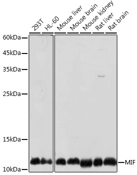
Western blot analysis of various lysates using MIF Rabbit pAb (CAB1391) at 1:1000 dilution. Secondary antibody: HRP-conjugated Goat anti-Rabbit IgG (H+L) (CABS014) at 1:10000 dilution. Lysates/proteins: 25μg per lane. Blocking buffer: 3% nonfat dry milk in TBST. Detection: ECL Basic Kit (AbGn00020). Exposure time: 10s.
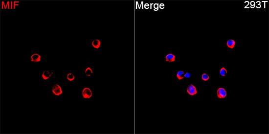
Immunofluorescence analysis of 293T cells using MIF Rabbit pAb(CAB1391) at a dilution of 1:100 (40x lens). Secondary antibody:Cy3 Goat Anti-Rabbit IgG (H+L)(CABS007) at 1:500 dilution. Blue: DAPI for nuclear staining.