The MIP Antibody (CAB2886) is a high-quality antibody developed for reliable detection and analysis of target proteins. Major intrinsic protein is a member of the water-transporting aquaporins as well as the original member of the MIP family of channel proteins. The function of the fiber cell membrane protein encoded by this gene is undetermined, yet this protein is speculated to play a role in intracellular communication. The MIP protein is expressed in the ocular lens and is required for correct lens function. This gene has been mapped among aquaporins AQP2, AQP5, and AQP6, in a potential gene cluster at 12q13.
This antibody is validated for use in WB, IHC-P, ELISA applications and has demonstrated reactivity against Human, Mouse, Rat samples.
Product Name:
MIP Antibody
SKU:
CAB2886
Size:
100μL, 20μL
Reactivity:
Human, Mouse, Rat
Conjugate:
Unconjugated
Immunogen:
Synthetic peptide. This information is considered to be commercially sensitive.
Tested Applications:
WBIHC-PELISA
Recommended Dilution:
WB
1:500 - 1:1000
IHC-P
1:100 - 1:200
ELISA
Recommended starting concentration is 1 μg/mL. Please optimize the concentration based on your specific assay requirements.
Cell Junction, Cell Membrane, Multi-Pass Membrane Protein, Gap Junction.
Calculated MW:
28kDa
Observed MW:
28kDa
Major intrinsic protein is a member of the water-transporting aquaporins as well as the original member of the MIP family of channel proteins. The function of the fiber cell membrane protein encoded by this gene is undetermined, yet this protein is speculated to play a role in intracellular communication. The MIP protein is expressed in the ocular lens and is required for correct lens function. This gene has been mapped among aquaporins AQP2, AQP5, and AQP6, in a potential gene cluster at 12q13.
Purification Method
Affinity purification
Gene ID
4284
RRID
AB_2764706
Buffer Information
Store at -20℃. Avoid freeze / thaw cycles. Buffer: PBS containing 50% glycerol, preserved with proclin300 or sodium azide, pH 7.3.
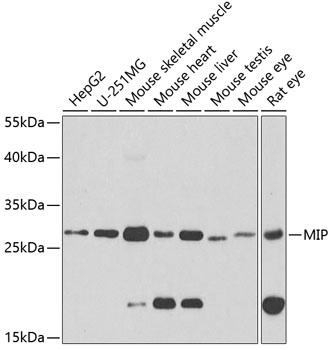
Western blot analysis of various lysates using MIP Rabbit pAb (CAB2886) at 1:1000 dilution. Secondary antibody: HRP-conjugated Goat anti-Rabbit IgG (H+L) (AS014) at 1:10000 dilution. Lysates/proteins: 25μg per lane. Blocking buffer: 3% nonfat dry milk in TBST. Detection: ECL Basic Kit (AbGn00020). Exposure time: 90s.
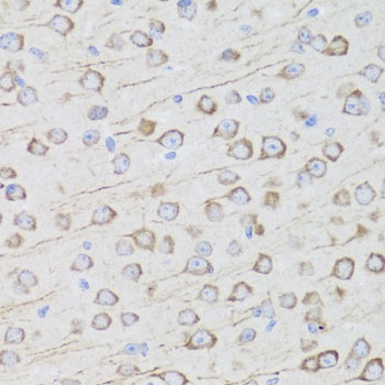
Immunohistochemistry analysis of paraffin-embedded Mouse brain using MIP Rabbit pAb (CAB2886) at dilution of 1:100 (40x lens). Microwave antigen retrieval performed with 0.01M PBS Buffer (pH 7.2) prior to IHC staining.
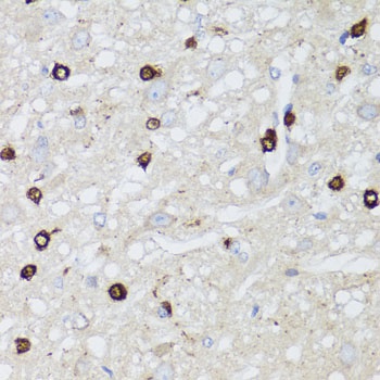
Immunohistochemistry analysis of paraffin-embedded Rat brain using MIP Rabbit pAb (CAB2886) at dilution of 1:100 (40x lens). Microwave antigen retrieval performed with 0.01M PBS Buffer (pH 7.2) prior to IHC staining.