The MITD1 Antibody (CAB18540) is a high-quality antibody developed for reliable detection and analysis of target proteins. This antibody, generated in rabbits, exhibits high reactivity with human samples and has been validated for use in Western blot applications. By binding specifically to the MITD1 protein, researchers can accurately detect and analyze MITD1 expression in a variety of cell types, making it an essential reagent for studies in cell biology and cancer research.
This antibody is validated for use in WB, IHC-P, ELISA applications and has demonstrated reactivity against Human, Mouse, Rat samples.
Product Name:
MITD1 Antibody
SKU:
CAB18540
Size:
20μL, 100μL
Reactivity:
Human, Mouse, Rat
Immunogen:
Recombinant protein (or fragment).This information is considered to be commercially sensitive.
Recommended starting concentration is 1 μg/mL. Please optimize the concentration based on your specific assay requirements.
Synonyms:
MITD1
Positive Sample:
U-87MG, MCF7, Mouse thymus, Mouse spleen, Rat thymus, Rat spleen
Cellular Localization:
Extracellular Exosome.
Calculated MW:
29kDa
Observed MW:
29kDa
Abscission, the separation of daughter cells at the end of cytokinesis, is effected by endosomal sorting complexes required for transport III (ESCRT-III). The protein encoded by this gene functions as a homodimer, with the N-termini binding to a subset of ESCRT-III subunits and the C-termini binding to membranes. The encoded protein regulates ESCRT-III activity and is required for proper cytokinesis. Several transcript variants encoding different isoforms have been found for this gene.
Purification Method
Affinity purification
Gene ID
129531
RRID
AB_2862304
Buffer Information
Store at -20℃. Avoid freeze / thaw cycles. Buffer: PBS with 0.01% thimerosal,50% glycerol,pH7.3.
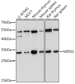
Western blot analysis of various lysates using MITD1 Rabbit pAb (CAB18540) at 1:1000 dilution. Secondary antibody: HRP-conjugated Goat anti-Rabbit IgG (H+L) (CABS014) at 1:10000 dilution. Lysates/proteins: 25μg per lane. Blocking buffer: 3% nonfat dry milk in TBST. Detection: ECL Enhanced Kit (AbGn00021). Exposure time: 180s.
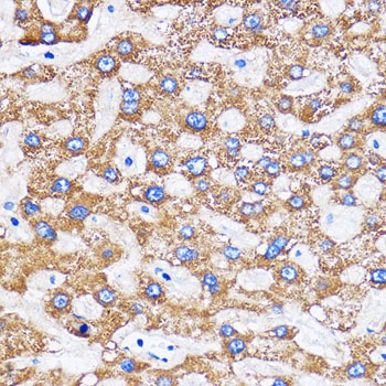
Immunohistochemistry analysis of paraffin-embedded Human liver using MITD1 Rabbit pAb (CAB18540) at dilution of 1:100 (40x lens). Microwave antigen retrieval performed with 0.01M PBS Buffer (pH 7.2) prior to IHC staining.