The MKNK1 Antibody (CAB18429) is a high-quality antibody developed for reliable detection and analysis of target proteins. This rabbit polyclonal antibody is highly reactive with human samples and has been validated for use in Western blot applications. By binding to the MKNK1 protein, this antibody enables accurate detection and analysis in various cell types, making it ideal for studies in cell signaling, cancer biology, and drug development.MKNK1 is a key player in the MAP kinase signaling pathway, which is known to regulate important cellular processes such as cell differentiation, proliferation, and survival.
This antibody is validated for use in WB, IHC-P, IF/ICC, ELISA applications and has demonstrated reactivity against Human, Mouse, Rat samples.
Product Name:
MKNK1 Antibody
SKU:
CAB18429
Size:
20μL, 100μL
Reactivity:
Human, Mouse, Rat
Immunogen:
Recombinant protein (or fragment).This information is considered to be commercially sensitive.
Recommended starting concentration is 1 μg/mL. Please optimize the concentration based on your specific assay requirements.
Synonyms:
MNK1, MKNK1
Positive Sample:
Mouse pancreas
Cellular Localization:
Cytoplasm, Cytoplasm, Nucleus.
Calculated MW:
51kDa
Observed MW:
51kDa
This gene encodes a Ser/Thr protein kinase that interacts with, and is activated by ERK1 and p38 mitogen-activated protein kinases, and thus may play a role in the response to environmental stress and cytokines. This kinase may also regulate transcription by phosphorylating eIF4E via interaction with the C-terminal region of eIF4G. Alternatively spliced transcript variants have been noted for this gene.
Purification Method
Affinity purification
Gene ID
8569
RRID
AB_2862196
Buffer Information
Store at -20℃. Avoid freeze / thaw cycles. Buffer: PBS containing 50% glycerol, preserved with proclin300 or sodium azide, pH 7.3.
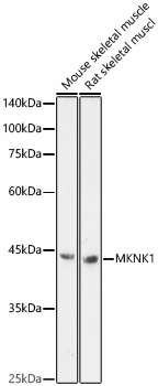
Western blot analysis of various lysates, using MKNK1 Rabbit pAb (CAB18429) at 1:1000 dilution. Secondary antibody: HRP-conjugated Goat anti-Rabbit IgG (H+L) (CABS014) at 1:10000 dilution. Lysates/proteins: 25μg per lane. Blocking buffer: 3% nonfat dry milk in TBST. Detection: ECL Enhanced Kit (AbGn00021). Exposure time: 180s.
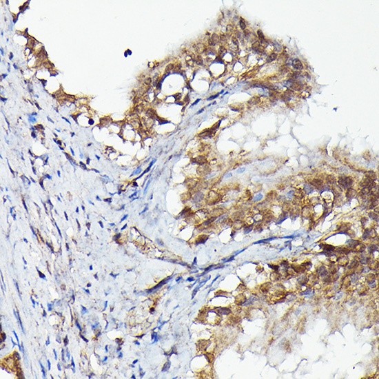
Immunohistochemistry analysis of paraffin-embedded Rat fallopian tube using MKNK1 Rabbit pAb (CAB18429) at dilution of 1:50 (40x lens). High pressure antigen retrieval performed with 0.01M Citrate buffer (pH 6.0) prior to IHC staining.
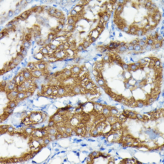
Immunohistochemistry analysis of paraffin-embedded Mouse kidney using MKNK1 Rabbit pAb (CAB18429) at dilution of 1:50 (40x lens). High pressure antigen retrieval performed with 0.01M Citrate buffer (pH 6.0) prior to IHC staining.
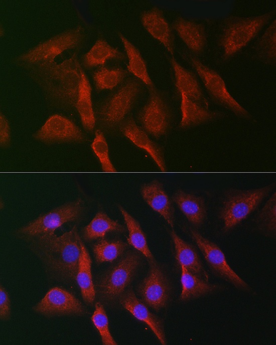
Immunofluorescence analysis of NIH-3T3 cells using MKNK1 Rabbit pAb (CAB18429) at dilution of 1:50 (40x lens). Secondary antibody: Cy3-conjugated Goat anti-Rabbit IgG (H+L) (CABS007) at 1:500 dilution. Blue: DAPI for nuclear staining.