The MOB1A/B Monoclonal Antibody (CAB23963) is a high-quality antibody developed for reliable detection and analysis of target proteins. These proteins are key regulators of cell proliferation and cell cycle progression, making them important targets for study in cancer research and developmental biology.This monoclonal antibody, produced through hybridoma technology, offers high specificity and sensitivity in detecting MOB1A and MOB1B proteins in Western blot and immunofluorescence applications.
This antibody is validated for use in WB, ELISA applications and has demonstrated reactivity against Human, Mouse, Rat samples.
Product Name:
MOB1A/B Monoclonal Antibody
SKU:
CAB23963
Size:
20μL, 100μL
Reactivity:
Human, Mouse, Rat
Clone Number:
ARC62859
Conjugate:
Unconjugated
Immunogen:
Synthetic peptide. This information is considered to be commercially sensitive.
The protein encoded by this gene is a component of the Hippo signaling pathway, which controls organ size and tumor growth by enhancing apoptosis. Loss of the encoded protein results in cell proliferation and cancer formation. The encoded protein is also involved in the control of microtubule stability during cytokinesis. Several transcript variants encoding different isoforms have been found for this gene.
Purification Method
Affinity purification
Gene ID
55233 92597
Buffer Information
Store at -20℃. Avoid freeze / thaw cycles. Buffer: PBS containing 50% glycerol and 0.05% BSA, preserved with proclin300 or sodium azide, pH 7.3.
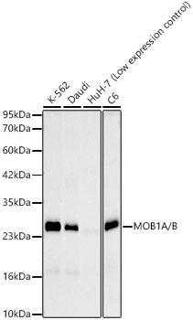
Western blot analysis of various lysates, using MOB1A/B Rabbit mAb (CAB23963) at 1:3000 dilution dilution incubated overnight at 4℃. Secondary antibody: HRP-conjugated Goat anti-Rabbit IgG (H+L) (CABS014) at 1:10000 dilution. Lysates/proteins: 25 μg per lane. Blocking buffer: 3% nonfat dry milk in TBST. Detection: ECL Basic Kit (AbGn00020). Exposure time: 30 s.
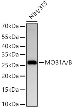
Western blot analysis of lysates from NIH/3T3 cells using MOB1A/B Rabbit mAb (CAB23963) at 1:2000 dilution incubated overnight at 4℃. Secondary antibody: HRP-conjugated Goat anti-Rabbit IgG (H+L) (CABS014) at 1:10000 dilution. Lysates/proteins: 25 μg per lane. Blocking buffer: 3% nonfat dry milk in TBST. Detection: ECL Basic Kit (AbGn00020). Exposure time: 45 s.
 at 1:3000 dilution. Secondary antibody: HRP Goat Anti-Rabbit IgG (H+L) at 1:10000 dilution. Lysates/proteins: 25ug per lane. Blocking buffer: 3% nonfat dry milk in TBST.")
 at 1:3000 dilution. Secondary antibody: HRP Goat Anti-Rabbit IgG (H+L) at 1:10000 dilution. Lysates/proteins: 25ug per lane. Blocking buffer: 3% nonfat dry milk in TBST.")


