The Mouse anti HA-Tag Monclonal Antibody (CABE008) is a high-quality antibody developed for reliable detection and analysis of target proteins. The HA-tag (YPYDVPDYA) is a short epitope derived from influenza hemagglutinin that is genetically incorporated into recombinant proteins to provide a specific immunological handle for detection and purification. It is particularly well-suited for western blotting, immunofluorescence, and immunoprecipitation due to the high affinity and specificity of anti-HA antibodies.
This antibody is validated for use in WB, IF/ICC, IP, ELISA applications and has demonstrated reactivity against Species independent samples.
Product Name:
Mouse anti HA-Tag Monclonal Antibody
SKU:
CABE008
Size:
20μL, 50μL, 100μL
Reactivity:
Species independent
Clone Number:
AMC0503
Conjugate:
Unconjugated
Immunogen:
Synthetic peptide. This information is considered to be commercially sensitive.
Tested Applications:
WBIF/ICCIPELISA
Recommended Dilution:
WB
1:2000 - 1:20000
IF/ICC
1:50 - 1:200
IP
0.5μg-4μg antibody for 200μg-400μg extracts of whole cells
ELISA
Recommended starting concentration is 1 μg/mL. Please optimize the concentration based on your specific assay requirements.
Synonyms:
HA, HA tag, HA-tag
Positive Sample:
293T transfected with HA-tag
Observed MW:
RefertoFigures
Protein tags are peptide sequences genetically grafted onto a recombinant protein. Often these tags are removable by chemical agents or by enzymatic means, such as proteolysis or intein splicing. Tags are attached to proteins for various purposes.Epitope tags are short peptide sequences which are chosen because high-affinity antibodies can be reliably produced in many different species. These are usually derived from viral genes, which explain their high immunoreactivity. Epitope tags include V5-tag, Myc-tag, HA-tag and NE-tag. These tags are particularly useful for western blotting, immunofluorescence and immunoprecipitation experiments, although they also find use in antibody purification.
Purification Method
Affinity purification
RRID
AB_2770404
Buffer Information
Store at -20℃. Avoid freeze / thaw cycles. Buffer: PBS containing 50% glycerol, preserved with proclin300 or sodium azide, pH 7.3.
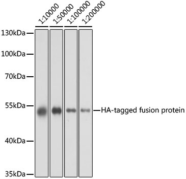
Western blot analysis of over-expressed HA-tagged protein in 293T cell using Mouse anti HA-Tag mAb (CABE008) at different dilution. Each lane was loaded with 2 ug cell lysate. Secondary antibody: HRP-conjugated Goat anti-Mouse IgG (H+L) (CABS003) at 1:10000 dilution. Blocking buffer: 3% nonfat dry milk in TBST. Detection: ECL Basic Kit (AbGn00020). Exposure time: 1s.
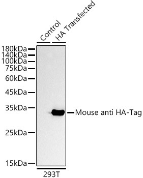
Western blot analysis of lysates from 293T cells, using Mouse anti HA-Tag mAb (CABE008) at 1:5000 dilution. Secondary antibody: HRP-conjugated Goat anti-Mouse IgG (H+L) (CABS003) at 1:10000 dilution. Lysates/proteins: 25μg per lane. Blocking buffer: 3% nonfat dry milk in TBST. Detection: ECL Basic Kit (AbGn00020). Exposure time: 10s.
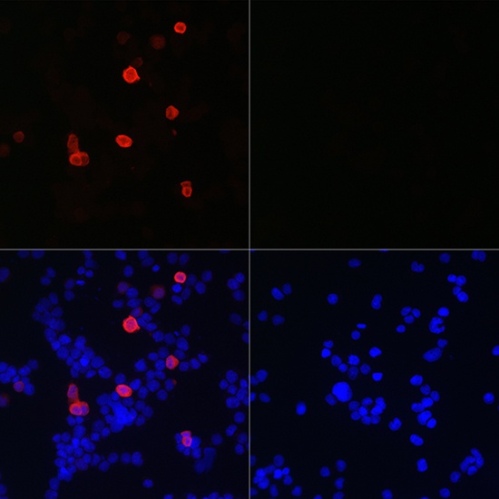
Immunofluorescence analysis of 293T cells transfected with HA-Tag fusion protein and untreated 293T cells use Mouse anti HA-Tag mAb (CABE008) at dilution of 1:50 (40x lens). Blue: DAPI for nuclear staining.
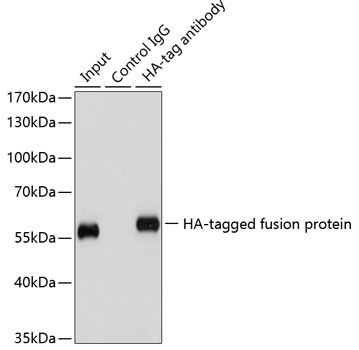
Immunoprecipitation of over-expressed HA-tagged protein in 293T cells using HA-tag antibody (CABE008). A mock served as negative control and over-expressed 293T cell lysate served as positive control.