The Mouse anti His-Tag Monclonal Antibody (CABE003) is a high-quality antibody developed for reliable detection and analysis of target proteins. The His-tag consists of consecutive histidine residues (typically 6–10) inserted into recombinant protein sequences, enabling detection and affinity purification via metal ion chelation on IMAC resins. His-tagged proteins can be efficiently captured from complex lysates under both native and denaturing conditions for downstream analysis.
This antibody is validated for use in WB, IF/ICC, IP, ELISA applications and has demonstrated reactivity against Species independent samples.
Product Name:
Mouse anti His-Tag Monclonal Antibody
SKU:
CABE003
Size:
20μL, 50μL, 100μL, 200μL
Reactivity:
Species independent
Clone Number:
AMC0149
Conjugate:
Unconjugated
Immunogen:
Synthetic peptide. This information is considered to be commercially sensitive.
Tested Applications:
WBIF/ICCIPELISA
Recommended Dilution:
WB
1:5000 - 1:30000
IF/ICC
1:5000-1:10000
IP
0.5ug-4ug antibody for 200ug-400ug extracts of whole cells
ELISA
Recommended starting concentration is 1 μg/mL. Please optimize the concentration based on your specific assay requirements.
Synonyms:
His, His tag, His-tag
Positive Sample:
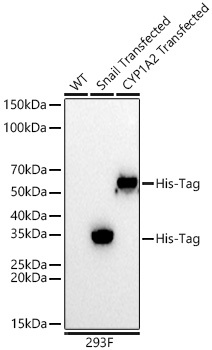
293F transfected with Snail-His-Tag, 293F transfected with CYP1A2-His-Tag
Observed MW:
35kDa(Snailtransfected)/58kDa(CYP1A2transfected)
Consecutive histidine residues (usually 6 to 10 in length) are often inserted into the amino acid sequences of recombinant proteins. The resulting His-tagged proteins can be detected by using His antibodies.
Purification Method
Affinity purification
RRID
AB_2728734
Buffer Information
Store at -20℃. Avoid freeze / thaw cycles. Buffer: PBS containing 50% glycerol, preserved with proclin300 or sodium azide, pH 7.3.
Western blot analysis of lysates from wild type (WT) and 293F cells transfected with His-Tag using Mouse anti His-Tag mAb (CABE003) at 1:30000 dilution incubated overnight at 4℃. Secondary antibody: HRP-conjugated Goat anti-Mouse IgG (H+L) (CABS003) at 1:10000 dilution. Lysates/proteins: 20 μg per lane. Blocking buffer: 3% nonfat dry milk in TBST. Detection: ECL Basic Kit (AbGn00020) .Exposure time: 5 s.
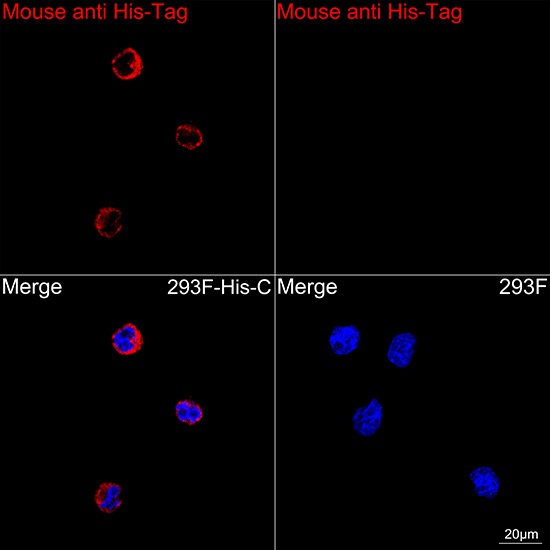
Confocal imaging of 293F cells transfected with His-C using Mouse anti His-Tag mAb (CABE003, dilution 1:10000) followed by a further incubation with Cy3 Goat Anti-Mouse IgG (H+L) (CABS008, dilution 1:500) (Red). DAPI was used for nuclear staining (Blue). Objective: 100x.
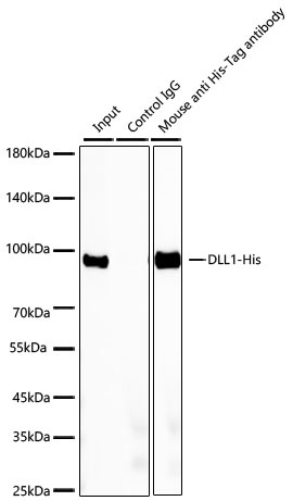
Immunoprecipitation of DLL1-His from 300 µg extracts of 293F cells transfected with a DLL1 expression vector containing a single N-terminal His-Tag was performed using 1 µg of Mouse anti His-Tag mAb (CABE003). Mouse IgG isotype control (AC011) was used to precipitate the Control IgG sample. The IP sample was eluted with 1X reducing Laemmli Buffer. The Input lane represents 10% of the total input. Western blot analysis of immunoprecipitates was conducted using Rabbit anti His-tag mAb (AE086) at a dilution of 1:10000.