The MRI1 Antibody (CAB14344) is a high-quality antibody developed for reliable detection and analysis of target proteins. This antibody, produced in rabbits, exhibits high specificity and sensitivity towards human samples, making it an ideal choice for Western blot applications. By binding to the MRI1 protein, this antibody enables the detection and analysis of MRI1 in various cell types, providing valuable insights into its role in cellular processes related to energy production and metabolism.MRI1, also known as N-methylpyristoyl transferase 1, is essential for the biogenesis of mitochondrial ribosomes and plays a crucial role in mitochondrial translation.
This antibody is validated for use in WB, ELISA applications and has demonstrated reactivity against Human, Mouse, Rat samples.
Product Name:
MRI1 Antibody
SKU:
CAB14344
Size:
20μL, 100μL
Reactivity:
Human, Mouse, Rat
Conjugate:
Unconjugated
Immunogen:
Recombinant protein (or fragment).This information is considered to be commercially sensitive.
Recommended starting concentration is 1 μg/mL. Please optimize the concentration based on your specific assay requirements.
Synonyms:
M1Pi, MRDI, MTNA, Ypr118w, MRI1
Positive Sample:
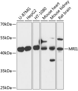
U-87MG, HepG2, HT-1080, Mouse heart, Mouse kidney, Rat brain
Cellular Localization:
Cell Projection, Cytoplasm, Nucleus.
Calculated MW:
39kDa
Observed MW:
45kDa
This enzyme functions in the methionine salvage pathway by catalyzing the interconversion of methylthioribose-1-phosphate and methythioribulose-1-phosphate. Elevated expression of the encoded protein is associated with metastatic melanoma and this protein promotes melanoma cell invasion independent of its enzymatic activity. Mutations in this gene may be associated with vanishing white matter disease (VMWD).
Purification Method
Affinity purification
Gene ID
84245
RRID
AB_2761210
Buffer Information
Store at -20℃. Avoid freeze / thaw cycles. Buffer: PBS with 0.01% thimerosal,50% glycerol,pH7.3.
Western blot analysis of various lysates using MRI1 Rabbit pAb (CAB14344) at 1:3000 dilution. Secondary antibody: HRP-conjugated Goat anti-Rabbit IgG (H+L) (CABS014) at 1:10000 dilution. Lysates/proteins: 25μg per lane. Blocking buffer: 3% nonfat dry milk in TBST. Detection: ECL Basic Kit (AbGn00020). Exposure time: 1s.