The MRPL42 Antibody (CAB12811) is a high-quality antibody developed for reliable detection and analysis of target proteins. This antibody, produced in rabbits, is highly specific to human samples and has been validated for use in various applications, including Western blot and immunofluorescence.MRPL42 is essential for the proper functioning of mitochondria, the powerhouse of the cell, and is crucial for maintaining cellular energy production. Dysregulation of MRPL42 has been linked to various diseases, including cancer and metabolic disorders, making it a promising target for further research and potential therapeutic interventions.
This antibody is validated for use in WB, ELISA applications and has demonstrated reactivity against Human, Mouse samples.
Product Name:
MRPL42 Antibody
SKU:
CAB12811
Size:
20μL, 100μL
Reactivity:
Human, Mouse
Conjugate:
Unconjugated
Immunogen:
Recombinant protein (or fragment).This information is considered to be commercially sensitive.
Mammalian mitochondrial ribosomal proteins are encoded by nuclear genes and help in protein synthesis within the mitochondrion. Mitochondrial ribosomes (mitoribosomes) consist of a small 28S subunit and a large 39S subunit. They have an estimated 75% protein to rRNA composition compared to prokaryotic ribosomes, where this ratio is reversed. Another difference between mammalian mitoribosomes and prokaryotic ribosomes is that the latter contain a 5S rRNA. Among different species, the proteins comprising the mitoribosome differ greatly in sequence, and sometimes in biochemical properties, which prevents easy recognition by sequence homology. This gene encodes a protein identified as belonging to both the 28S and the 39S subunits. Alternative splicing results in multiple transcript variants. Pseudogenes corresponding to this gene are found on chromosomes 4q, 6p, 6q, 7p, and 15q.
Purification Method
Affinity purification
Gene ID
28977
RRID
AB_2759651
Buffer Information
Store at -20℃. Avoid freeze / thaw cycles. Buffer: PBS with 0.01% thimerosal,50% glycerol,pH7.3.
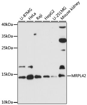
Western blot analysis of various lysates using MRPL42 Rabbit pAb (CAB12811) at 1:3000 dilution. Secondary antibody: HRP-conjugated Goat anti-Rabbit IgG (H+L) (CABS014) at 1:10000 dilution. Lysates/proteins: 25μg per lane. Blocking buffer: 3% nonfat dry milk in TBST. Detection: ECL Basic Kit (AbGn00020). Exposure time: 90s.