The MRPL54 Antibody (CAB14957) is a high-quality antibody developed for reliable detection and analysis of target proteins. This antibody, produced in rabbits, exhibits high reactivity with human samples and is validated for use in Western blotting applications. By binding specifically to the MRPL54 protein, researchers can accurately detect and analyze its expression in various cell types, making it ideal for investigations in mitochondrial biology and related fields.MRPL54 is a key component of the mitochondrial ribosome complex that is essential for the translation of mitochondrial DNA-encoded proteins.
This antibody is validated for use in WB, IHC-P, ELISA applications and has demonstrated reactivity against Human samples.
Product Name:
MRPL54 Antibody
SKU:
CAB14957
Size:
20μL, 100μL
Reactivity:
Human
Conjugate:
Unconjugated
Immunogen:
Recombinant protein (or fragment).This information is considered to be commercially sensitive.
Recommended starting concentration is 1 μg/mL. Please optimize the concentration based on your specific assay requirements.
Synonyms:
L54mt, MRP-L54, MRPL54
Positive Sample:
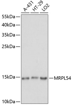
A-431, HT-29, LO2
Cellular Localization:
Mitochondrion.
Calculated MW:
16kDa
Observed MW:
15kDa
Mammalian mitochondrial ribosomal proteins are encoded by nuclear genes and help in protein synthesis within the mitochondrion. Mitochondrial ribosomes (mitoribosomes) consist of a small 28S subunit and a large 39S subunit. They have an estimated 75% protein to rRNA composition compared to prokaryotic ribosomes, where this ratio is reversed. Another difference between mammalian mitoribosomes and prokaryotic ribosomes is that the latter contain a 5S rRNA. Among different species, the proteins comprising the mitoribosome differ greatly in sequence, and sometimes in biochemical properties, which prevents easy recognition by sequence homology. This gene encodes a 39S subunit protein.
Purification Method
Affinity purification
Gene ID
116541
RRID
AB_2761840
Buffer Information
Store at -20℃. Avoid freeze / thaw cycles. Buffer: PBS with 0.01% thimerosal,50% glycerol,pH7.3.
Western blot analysis of various lysates using MRPL54 Rabbit pAb (CAB14957) at 1:1000 dilution. Secondary antibody: HRP-conjugated Goat anti-Rabbit IgG (H+L) (CABS014) at 1:10000 dilution. Lysates/proteins: 25μg per lane. Blocking buffer: 3% nonfat dry milk in TBST. Detection: ECL Basic Kit (AbGn00020). Exposure time: 30s.
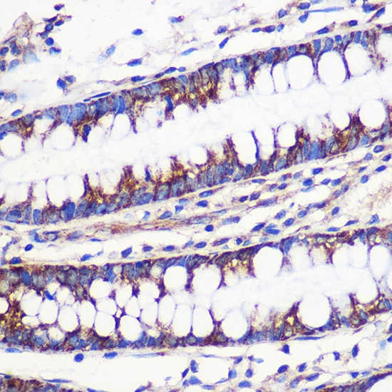
Immunohistochemistry analysis of paraffin-embedded Human colon using MRPL54 Rabbit pAb (CAB14957) at dilution of 1:100 (40x lens). Microwave antigen retrieval performed with 0.01M PBS Buffer (pH 7.2) prior to IHC staining.