The MSH4 Antibody (CAB8556) is a high-quality antibody developed for reliable detection and analysis of target proteins. This antibody, produced in rabbits, has high specificity and sensitivity for detecting MSH4 in human samples, making it ideal for use in Western blot applications.MSH4 is a member of the MutS homolog family and plays a crucial role in the process of meiotic recombination, ensuring the accurate segregation of chromosomes during cell division. Dysregulation of MSH4 has been implicated in genetic disorders and tumorigenesis, making it a promising target for cancer research and understanding genetic diseases.
This antibody is validated for use in WB, ELISA applications and has demonstrated reactivity against Mouse, Rat samples.
Product Name:
MSH4 Antibody
SKU:
CAB8556
Size:
20μL, 100μL
Reactivity:
Mouse, Rat
Conjugate:
Unconjugated
Immunogen:
Recombinant protein (or fragment).This information is considered to be commercially sensitive.
Recommended starting concentration is 1 μg/mL. Please optimize the concentration based on your specific assay requirements.
Synonyms:
ASG, POF20, SPGF2, MSH4
Positive Sample:
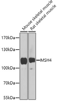
Mouse skeletal muscle, Rat skeletal muscle
Cellular Localization:
Nucleus.
Calculated MW:
105kDa
Observed MW:
105kDa
This gene encodes a member of the DNA mismatch repair mutS family. This member is a meiosis-specific protein that is not involved in DNA mismatch correction, but is required for reciprocal recombination and proper segregation of homologous chromosomes at meiosis I. This protein and MSH5 form a heterodimer which binds uniquely to a Holliday Junction and its developmental progenitor, thus provoking ADP-ATP exchange, and stabilizing the interaction between parental chromosomes during meiosis double-stranded break repair.
Purification Method
Affinity purification
Gene ID
4438
RRID
AB_2770446
Buffer Information
Store at -20℃. Avoid freeze / thaw cycles. Buffer: PBS containing 50% glycerol, preserved with proclin300 or sodium azide, pH 7.3.
Western blot analysis of various lysates using MSH4 Rabbit pAb (CAB8556) at 1:1000 dilution. Secondary antibody: HRP-conjugated Goat anti-Rabbit IgG (H+L) (CABS014) at 1:10000 dilution. Lysates/proteins: 25μg per lane. Blocking buffer: 3% nonfat dry milk in TBST. Detection: ECL Enhanced Kit (AbGn00021). Exposure time: 90s.