The MSRA Antibody (CAB6389) is a high-quality antibody developed for reliable detection and analysis of target proteins. MSRA is an antioxidant enzyme that repairs oxidized proteins, playing a key role in protecting cells from damage caused by reactive oxygen species.Raised in rabbits, this antibody is highly specific and sensitive for detecting MSRA in human samples. It has been validated for use in Western blot applications, enabling researchers to analyze MSRA expression levels in various cell types and tissues. This antibody binds specifically to the MSRA protein, allowing for precise detection and analysis in experimental studies.
This antibody is validated for use in WB, IHC-P, IF/ICC, ELISA applications and has demonstrated reactivity against Human, Mouse, Rat samples.
Product Name:
MSRA Antibody
SKU:
CAB6389
Size:
20μL, 100μL
Reactivity:
Human, Mouse, Rat
Conjugate:
Unconjugated
Immunogen:
Recombinant protein (or fragment).This information is considered to be commercially sensitive.
This gene encodes a ubiquitous and highly conserved protein that carries out the enzymatic reduction of methionine sulfoxide to methionine. Human and animal studies have shown the highest levels of expression in kidney and nervous tissue. The protein functions in the repair of oxidatively damaged proteins to restore biological activity. Alternative splicing results in multiple transcript variants.
Purification Method
Affinity purification
Gene ID
4482
RRID
AB_2766991
Buffer Information
Store at -20℃. Avoid freeze / thaw cycles. Buffer: PBS containing 50% glycerol, preserved with proclin300 or sodium azide, pH 7.3.
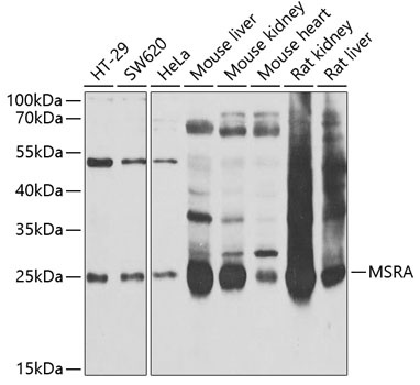
Western blot analysis of various lysates using MSRA Rabbit pAb (CAB6389) at 1:1000 dilution. Secondary antibody: HRP-conjugated Goat anti-Rabbit IgG (H+L) (CABS014) at 1:10000 dilution. Lysates/proteins: 25μg per lane. Blocking buffer: 3% nonfat dry milk in TBST. Detection: ECL Basic Kit (AbGn00020). Exposure time: 30s.
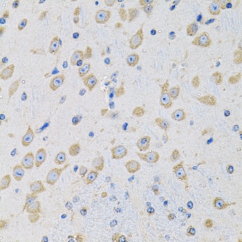
Immunohistochemistry analysis of paraffin-embedded Mouse brain using MSRA Rabbit pAb (CAB6389) at dilution of 1:100 (40x lens). Microwave antigen retrieval performed with 0.01M PBS Buffer (pH 7.2) prior to IHC staining.
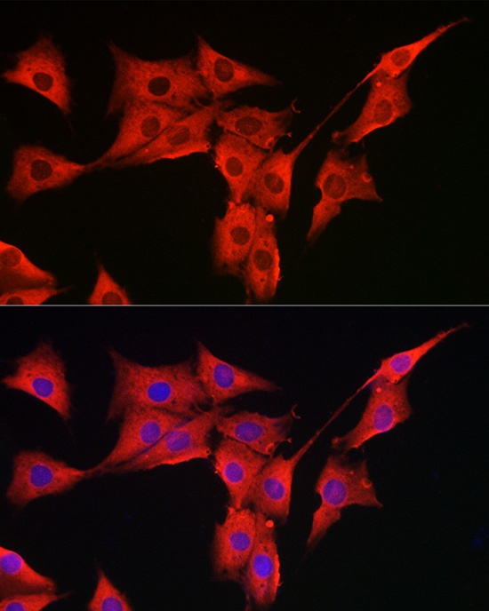
Immunofluorescence analysis of PC-12 cells using MSRA Rabbit pAb (CAB6389) at dilution of 1:300 (40x lens). Secondary antibody: Cy3-conjugated Goat anti-Rabbit IgG (H+L) (CABS007) at 1:500 dilution. Blue: DAPI for nuclear staining.
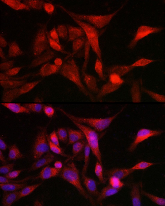
Immunofluorescence analysis of U2OS cells using MSRA Rabbit pAb (CAB6389) at dilution of 1:300 (40x lens). Secondary antibody: Cy3-conjugated Goat anti-Rabbit IgG (H+L) (CABS007) at 1:500 dilution. Blue: DAPI for nuclear staining.