The MT-ND1 Antibody (CAB17967) is a high-quality antibody developed for reliable detection and analysis of target proteins. This antibody, derived from rabbit polyclonal sources, is highly specific and reactive with human samples, making it ideal for use in Western blot applications. By binding to the MT-ND1 protein, researchers can detect and analyze its expression in various cell types, providing valuable insights into mitochondrial function and dysfunction.MT-ND1 is a key component of complex I in the mitochondrial electron transport chain, playing a crucial role in ATP production and cellular metabolism.
This antibody is validated for use in WB, IHC-P, IF/ICC, ELISA applications and has demonstrated reactivity against Human, Mouse, Rat samples.
Product Name:
MT-ND1 Antibody
SKU:
CAB17967
Size:
20μL, 100μL
Reactivity:
Human, Mouse, Rat
Immunogen:
Synthetic peptide. This information is considered to be commercially sensitive.
Recommended starting concentration is 1 μg/mL. Please optimize the concentration based on your specific assay requirements.
Synonyms:
MTND1, ND1, MT-ND1
Positive Sample:
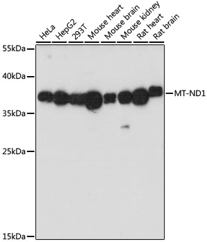
HeLa, HepG2, 293T, mouse heart, mouse brain, mouse kidney, rat heart, rat brain
Cellular Localization:
Mitochondrial Inner Membrane, Mitochondrial Membrane, Mitochondrial Respiratory Chain Complex I.
Calculated MW:
36kDa
Observed MW:
36kDa
Enables NADH dehydrogenase (ubiquinone) activity. Involved in mitochondrial electron transport, NADH to ubiquinone and mitochondrial respiratory chain complex I assembly. Located in mitochondrial membrane. Part of mitochondrial respiratory chain complex I. Implicated in several diseases, including MELAS syndrome; neurodegenerative disease (multiple); optic nerve disease (multiple); toxic shock syndrome; and type 2 diabetes mellitus. Biomarker of Alzheimer's disease; Parkinson's disease; and multiple sclerosis.
Purification Method
Affinity purification
Gene ID
4535
RRID
AB_2861769
Buffer Information
Store at -20℃. Avoid freeze / thaw cycles. Buffer: PBS with 0.09% Sodium azide,50% glycerol,pH7.3.
Western blot analysis of various lysates using MT-ND1 Rabbit pAb (CAB17967) at 1:1000 dilution. Secondary antibody: HRP-conjugated Goat anti-Rabbit IgG (H+L) (CABS014) at 1:10000 dilution. Lysates/proteins: 25μg per lane. Blocking buffer: 3% nonfat dry milk in TBST. Detection: ECL Basic Kit (AbGn00020). Exposure time: 90s.
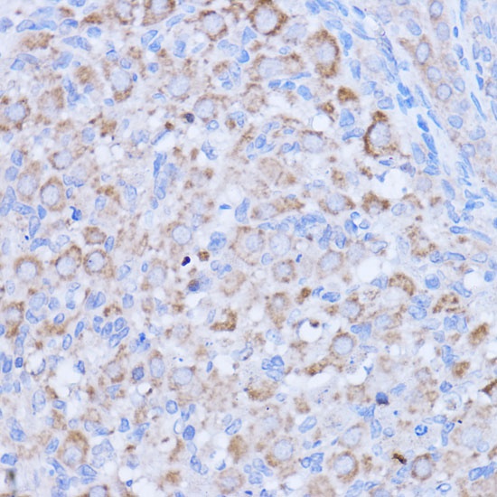
Immunohistochemistry analysis of paraffin-embedded Rat ovary using MT-ND1 Rabbit pAb (CAB17967) at dilution of 1:100 (40x lens). Microwave antigen retrieval performed with 0.01M PBS Buffer (pH 7.2) prior to IHC staining.
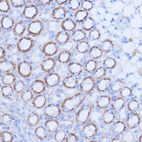
Immunohistochemistry analysis of paraffin-embedded Mouse kidney using MT-ND1 Rabbit pAb (CAB17967) at dilution of 1:100 (40x lens). Microwave antigen retrieval performed with 0.01M PBS Buffer (pH 7.2) prior to IHC staining.
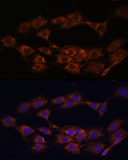
Immunofluorescence analysis of C6 cells using MT-ND1 Rabbit pAb (CAB17967) at dilution of 1:100 (40x lens). Secondary antibody: Cy3-conjugated Goat anti-Rabbit IgG (H+L) (CABS007) at 1:500 dilution. Blue: DAPI for nuclear staining.