The MTCH2 Antibody (CAB12934) is a high-quality antibody developed for reliable detection and analysis of target proteins. This antibody, generated in rabbits, exhibits high specificity and sensitivity towards human samples, allowing for accurate detection and analysis in Western blot experiments.MTCH2 is involved in various cellular processes, including mitochondrial fission, apoptosis, and metabolism regulation. Dysregulation of MTCH2 has been linked to diseases such as cancer, neurodegenerative disorders, and metabolic syndromes, highlighting its potential as a therapeutic target.
This antibody is validated for use in WB, IHC-P, IF/ICC, ELISA applications and has demonstrated reactivity against Human, Mouse samples.
Product Name:
MTCH2 Antibody
SKU:
CAB12934
Size:
20μL, 100μL
Reactivity:
Human, Mouse
Conjugate:
Unconjugated
Immunogen:
Recombinant protein (or fragment).This information is considered to be commercially sensitive.
This gene encodes a member of the SLC25 family of nuclear-encoded transporters that are localized in the inner mitochondrial membrane. Members of this superfamily are involved in many metabolic pathways and cell functions. Genome-wide association studies in human have identified single-nucleotide polymorphisms in several loci associated with obesity. This gene is one such locus, which is highly expressed in white adipose tissue and adipocytes, and thought to play a regulatory role in adipocyte differentiation and biology. Alternatively spliced transcript variants encoding different isoforms have been found for this gene. A recent study showed this gene to be an authentic stop codon readthrough target that can produce two isoforms from the same mRNA by use of alternative in-frame translation termination codons.
Purification Method
Affinity purification
Gene ID
23788
RRID
AB_2759780
Buffer Information
Store at -20℃. Avoid freeze / thaw cycles. Buffer: PBS with 0.01% thimerosal,50% glycerol,pH7.3.
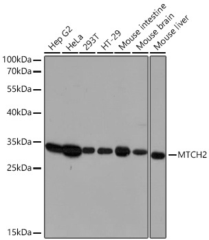
Western blot analysis of various lysates using [KO Validated] MTCH2 Rabbit pAb (CAB12934) at 1:3000 dilution incubated overnight at 4℃. Secondary antibody: HRP-conjugated Goat anti-Rabbit IgG (H+L) (CABS014) at 1:10000 dilution. Lysates/proteins: 25 μg per lane. Blocking buffer: 3% nonfat dry milk in TBST. Detection: ECL Basic Kit (AbGn00020). Exposure time: 30 s.
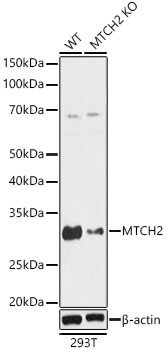
Western blot analysis of lysates from wild type(WT) and MTCH2 knockout (KO) HeLa(KO) cells, using [KO Validated] MTCH2 Rabbit pAb (CAB12934) at 1:1000 dilution incubated overnight at 4℃. Secondary antibody: HRP-conjugated Goat anti-Rabbit IgG (H+L) (CABS014) at 1:10000 dilution. Lysates/proteins: 25 μg per lane. Blocking buffer: 3% nonfat dry milk in TBST. Detection: ECL Basic Kit (AbGn00020). Exposure time: 3 s.
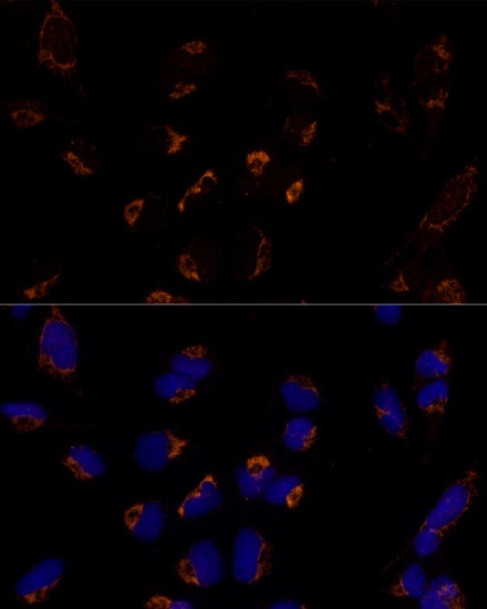
Immunofluorescence analysis of U-2 OS cells using [KO Validated] MTCH2 Rabbit pAb (CAB12934) at dilution of 1:100 (40x lens). Secondary antibody: Cy3-conjugated Goat anti-Rabbit IgG (H+L) (CABS007) at 1:500 dilution. Blue: DAPI for nuclear staining.