The MTHFD2 Monoclonal Antibody (CAB22653) is a high-quality antibody developed for reliable detection and analysis of target proteins. This monoclonal antibody, generated in mice, shows high specificity and sensitivity for detecting MTHFD2 in various cell types and tissues. It has been validated for use in applications such as immunohistochemistry and flow cytometry.MTHFD2 is a mitochondrial enzyme that plays a crucial role in one-carbon metabolism, which is essential for nucleotide biosynthesis and DNA methylation.
This antibody is validated for use in WB, ELISA applications and has demonstrated reactivity against Human, Mouse samples.
Product Name:
MTHFD2 Monoclonal Antibody
SKU:
CAB22653
Size:
20μL, 100μL
Reactivity:
Human, Mouse
Clone Number:
ARC58228
Conjugate:
Unconjugated
Immunogen:
Recombinant protein (or fragment).This information is considered to be commercially sensitive.
Recommended starting concentration is 1 μg/mL. Please optimize the concentration based on your specific assay requirements.
Synonyms:
NMDMC, MTHFD2
Positive Sample:
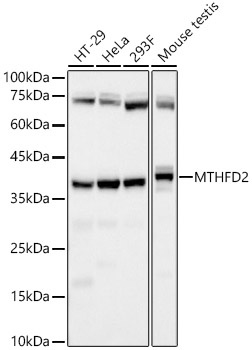
HT-29, HeLa, 293F, Mouse testis
Cellular Localization:
Mitochondrion.
Calculated MW:
38kDa
Observed MW:
Refertofigures
This gene encodes a nuclear-encoded mitochondrial bifunctional enzyme with methylenetetrahydrofolate dehydrogenase and methenyltetrahydrofolate cyclohydrolase activities. The enzyme functions as a homodimer and is unique in its absolute requirement for magnesium and inorganic phosphate. Formation of the enzyme-magnesium complex allows binding of NAD. Alternative splicing results in two different transcripts, one protein-coding and the other not protein-coding. This gene has a pseudogene on chromosome 7.
Purification Method
Affinity purification
Gene ID
10797
Buffer Information
Store at -20℃. Avoid freeze / thaw cycles. Buffer: PBS containing 50% glycerol and 0.05% BSA, preserved with proclin300 or sodium azide, pH 7.3.
Western blot analysis of various lysates, using MTHFD2 Rabbit mAb (CAB22653) at 1:7000 dilution. Secondary antibody: HRP-conjugated Goat anti-Rabbit IgG (H+L) (CABS014) at 1:10000 dilution. Lysates/proteins: 25μg per lane. Blocking buffer: 3% nonfat dry milk in TBST. Detection: ECL Basic Kit (AbGn00020). Exposure time: 30s.
 at 1:7000 dilution. Secondary antibody: HRP Goat Anti-Rabbit IgG (H+L) at 1:10000 dilution. Lysates/proteins: 25μg per lane. Blocking buffer: 3% nonfat dry milk in TBST.")
 at 1:7000 dilution. Secondary antibody: HRP Goat Anti-Rabbit IgG (H+L) at 1:10000 dilution. Lysates/proteins: 25μg per lane. Blocking buffer: 3% nonfat dry milk in TBST.")


")