The MUC1 Monoclonal Antibody (CAB19081) is a high-quality antibody developed for reliable detection and analysis of target proteins. This antibody, developed using rabbit monoclonal technology, exhibits high specificity and sensitivity towards human samples, making it ideal for use in Western blot and immunohistochemistry applications.MUC1 plays a pivotal role in cancer progression and metastasis by promoting tumor cell survival, proliferation, and immune evasion.
This antibody is validated for use in WB, IHC-P, ELISA applications and has demonstrated reactivity against Human, Mouse, Rat samples.
Product Name:
MUC1 Monoclonal Antibody
SKU:
CAB19081
Size:
20μL, 100μL
Reactivity:
Human, Mouse, Rat
Clone Number:
ARC0352
Conjugate:
Unconjugated
Immunogen:
Synthetic peptide. This information is considered to be commercially sensitive.
Apical Cell Membrane, Cell Membrane, Cytoplasm, Nucleus, Secreted, Single-Pass Type I Membrane Protein.
Calculated MW:
122kDa
Observed MW:
17-25kDa
This gene encodes a membrane-bound protein that is a member of the mucin family. Mucins are O-glycosylated proteins that play an essential role in forming protective mucous barriers on epithelial surfaces. These proteins also play a role in intracellular signaling. This protein is expressed on the apical surface of epithelial cells that line the mucosal surfaces of many different tissues including lung, breast stomach and pancreas. This protein is proteolytically cleaved into alpha and beta subunits that form a heterodimeric complex. The N-terminal alpha subunit functions in cell-adhesion and the C-terminal beta subunit is involved in cell signaling. Overexpression, aberrant intracellular localization, and changes in glycosylation of this protein have been associated with carcinomas. This gene is known to contain a highly polymorphic variable number tandem repeats (VNTR) domain. Alternate splicing results in multiple transcript variants.
Purification Method
Affinity purification
Gene ID
4582
RRID
AB_2862573
Buffer Information
Store at -20℃. Avoid freeze / thaw cycles. Buffer: PBS containing 50% glycerol and 0.05% BSA, preserved with proclin300 or sodium azide, pH 7.3.
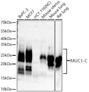
Western blot analysis of various lysates using MUC1-C Rabbit mAb (CAB19081) at 1:1000 dilution incubated at room temperature for 1.5 hours. Secondary antibody: HRP-conjugated Goat anti-Rabbit IgG (H+L) (CABS014) at 1:10000 dilution. Lysates/proteins: 25 μg per lane. Blocking buffer: 3% nonfat dry milk in TBST. Detection: ECL Basic Kit (AbGn00020). Negative control (NC): HCT 116. Exposure time: 45 s.
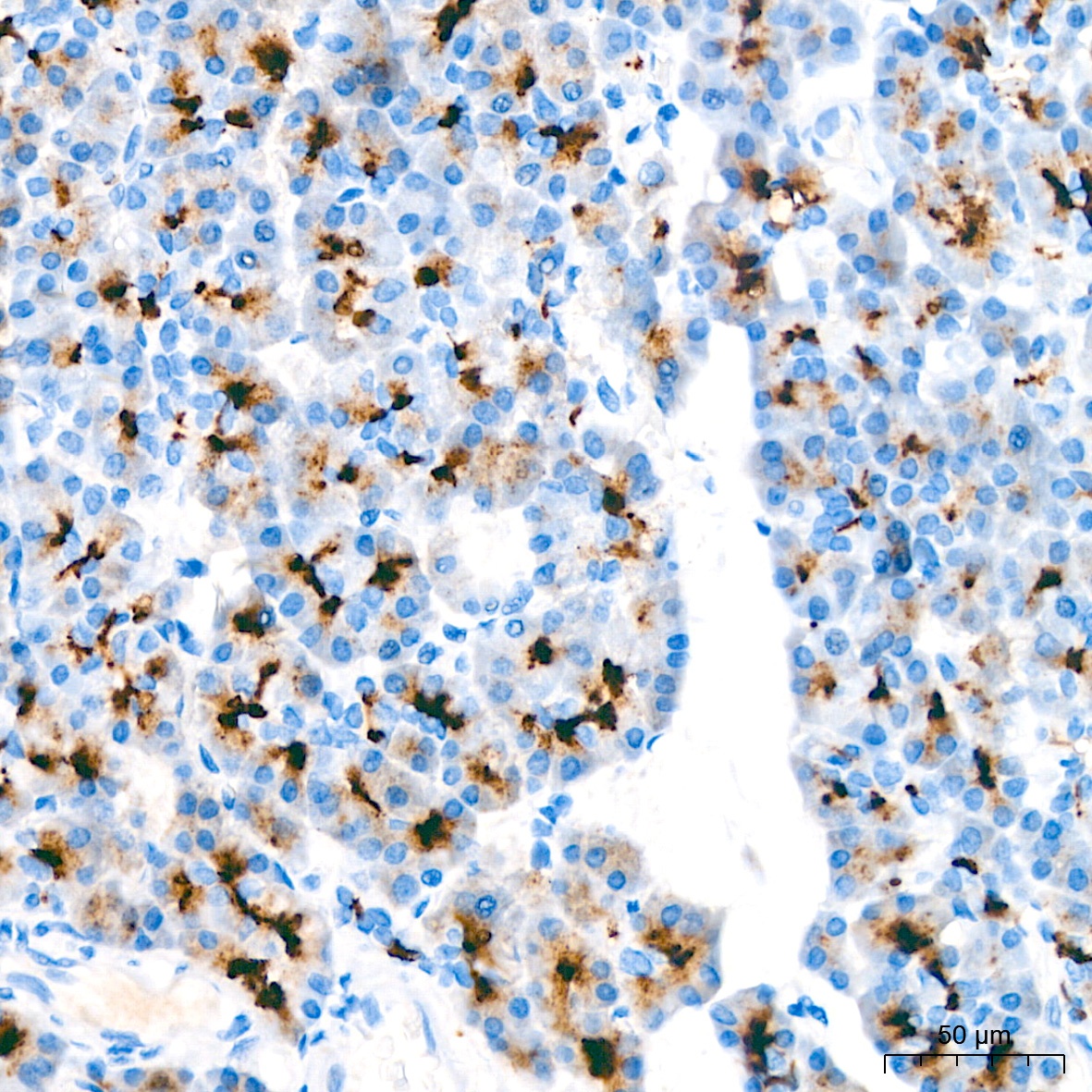
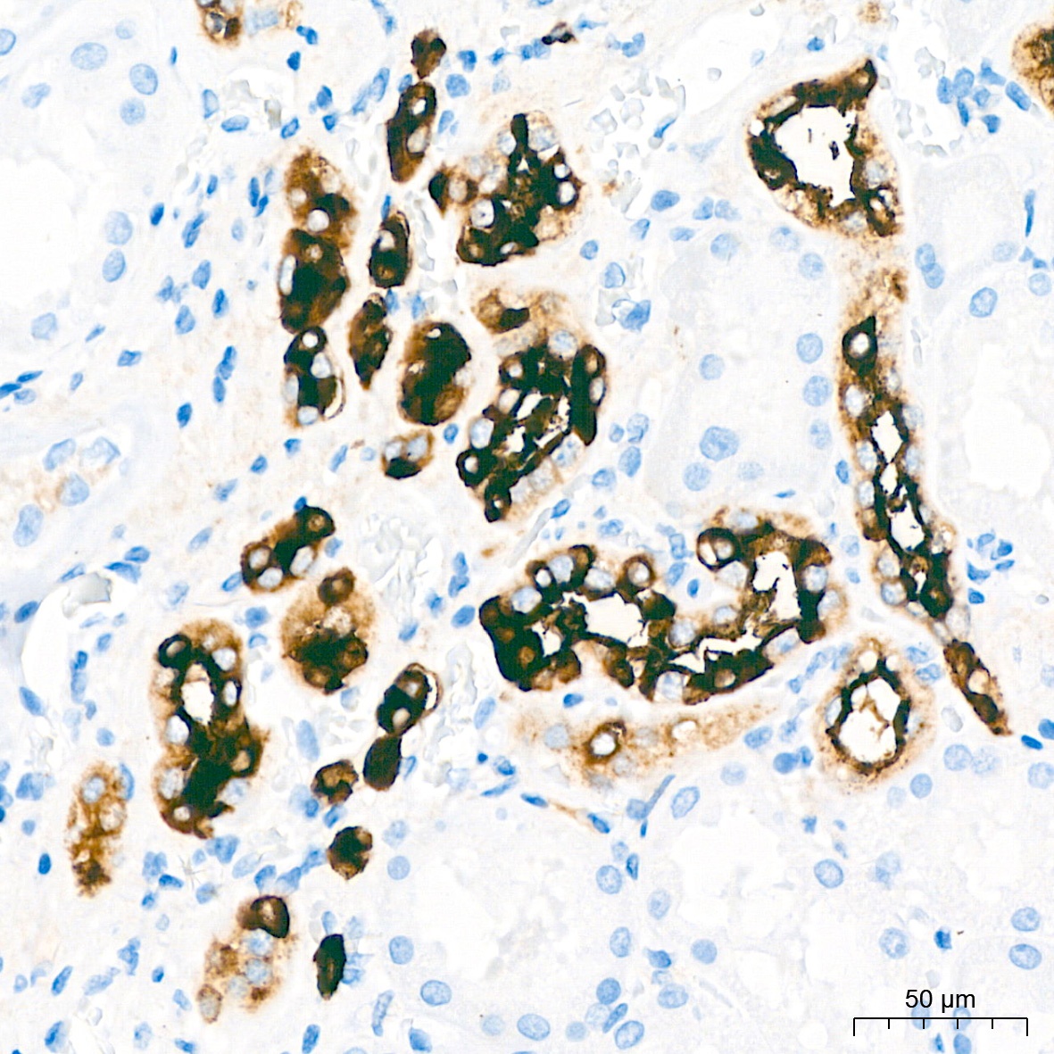
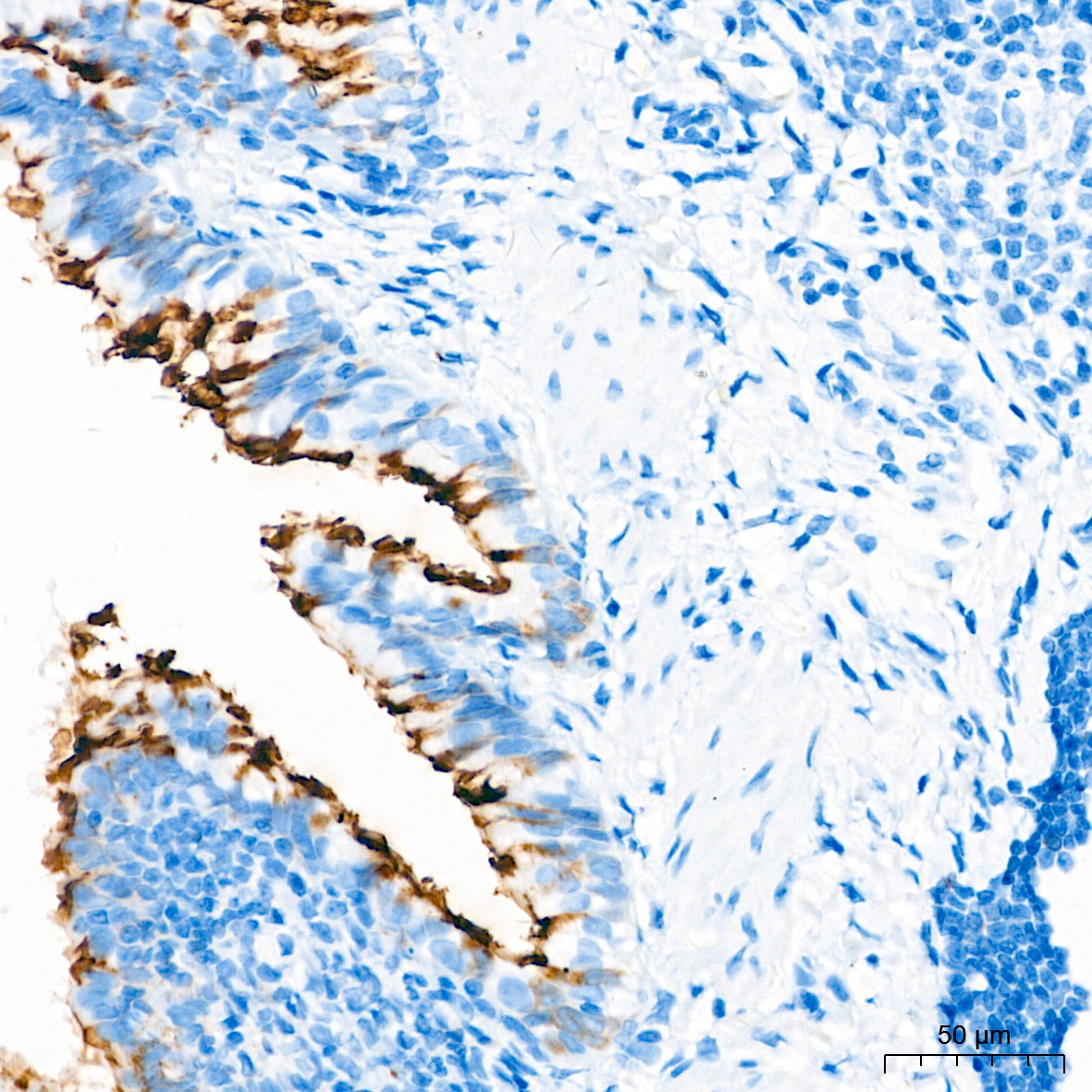
Immunohistochemistry analysis of paraffin-embedded Human pancreas tissue using MUC1-C Rabbit mAb (CAB19081) at a dilution of 1:400 (40x lens). High pressure antigen retrieval performed with 0.01M Tris-EDTA Buffer(pH 9.0) prior to IHC staining.
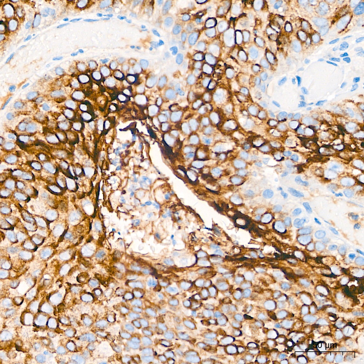
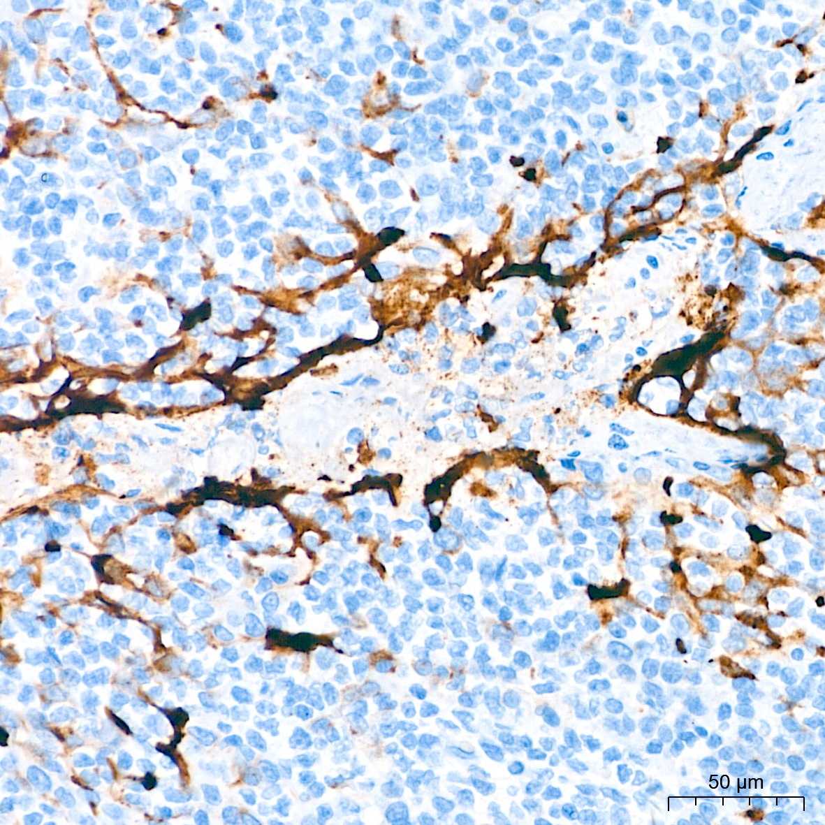
Immunohistochemistry analysis of paraffin-embedded Human cervix cancer tissue using MUC1-C Rabbit mAb (CAB19081) at a dilution of 1:400 (40x lens). High pressure antigen retrieval performed with 0.01M Tris-EDTA Buffer(pH 9.0) prior to IHC staining.
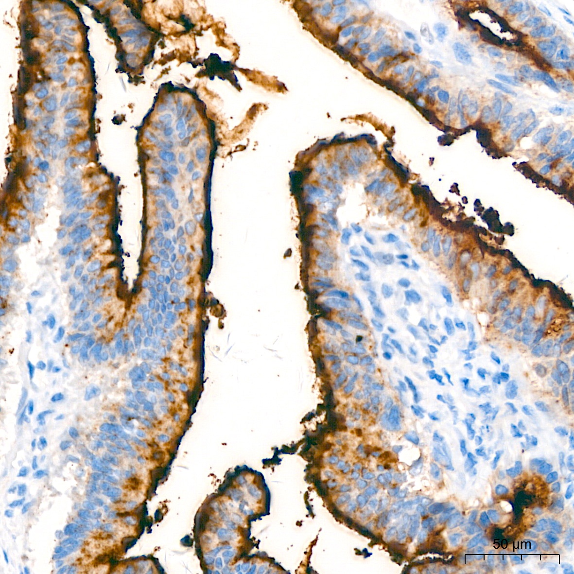
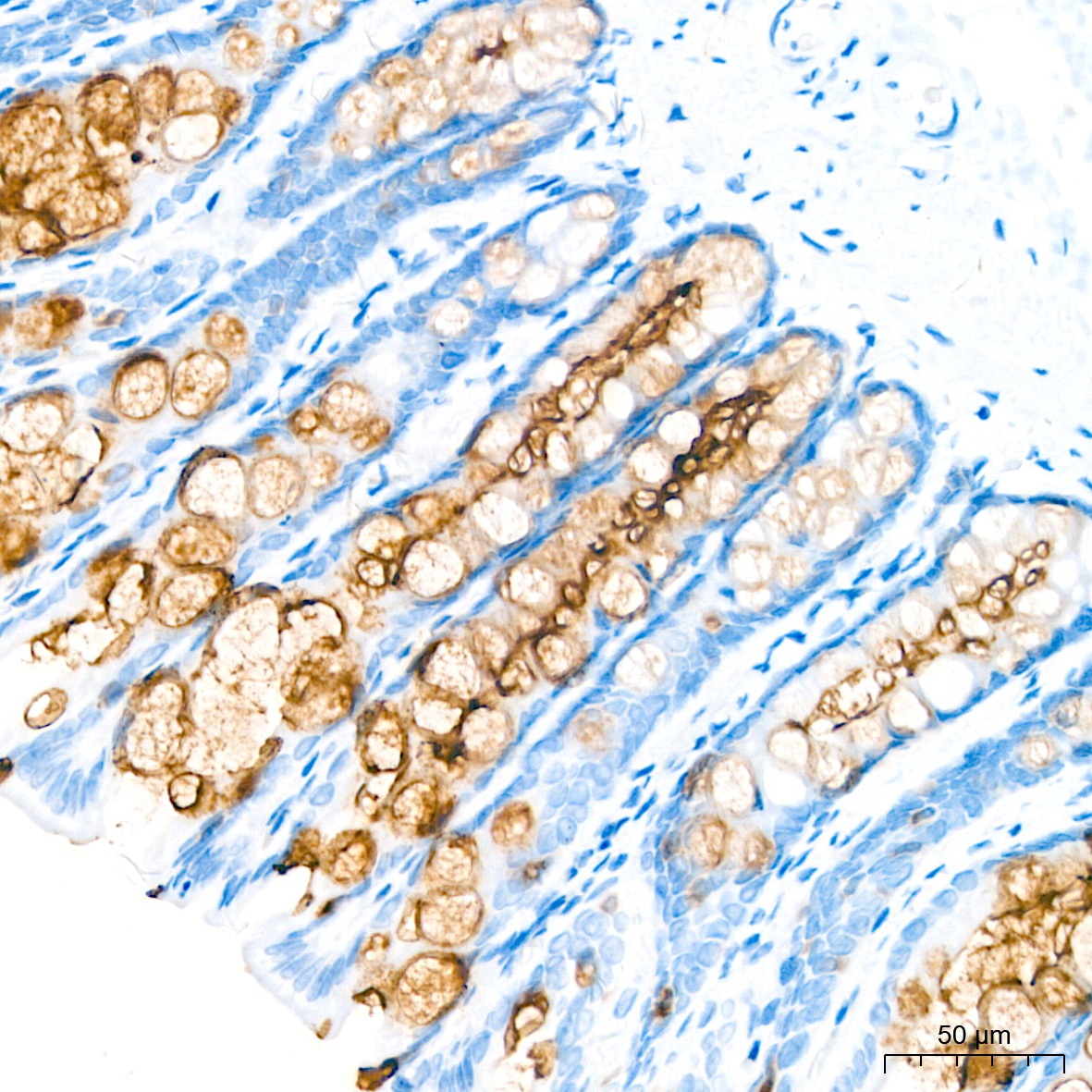
Immunohistochemistry analysis of paraffin-embedded Human colon carcinoma tissue using MUC1-C Rabbit mAb (CAB19081) at a dilution of 1:400 (40x lens). High pressure antigen retrieval performed with 0.01M Tris-EDTA Buffer(pH 9.0) prior to IHC staining.
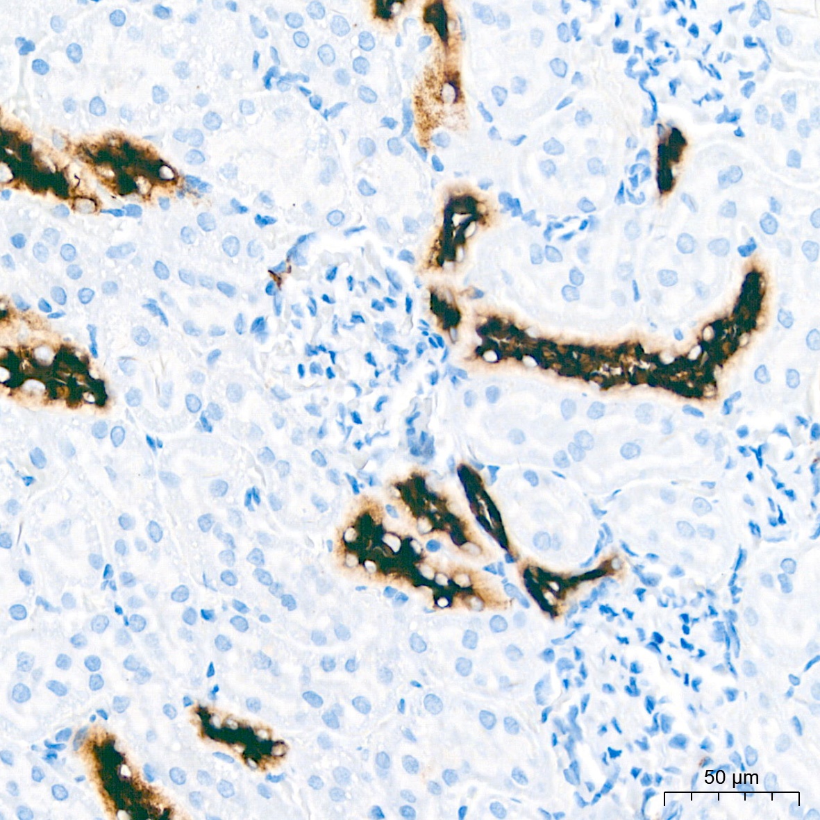
Immunohistochemistry analysis of paraffin-embedded Human kidney tissue using MUC1-C Rabbit mAb (CAB19081) at a dilution of 1:400 (40x lens). High pressure antigen retrieval performed with 0.01M Tris-EDTA Buffer(pH 9.0) prior to IHC staining.
Immunohistochemistry analysis of paraffin-embedded Human tonsil tissue using MUC1-C Rabbit mAb (CAB19081) at a dilution of 1:400 (40x lens). High pressure antigen retrieval performed with 0.01M Tris-EDTA Buffer(pH 9.0) prior to IHC staining.
Immunohistochemistry analysis of paraffin-embedded Rat colon tissue using MUC1-C Rabbit mAb (CAB19081) at a dilution of 1:400 (40x lens). High pressure antigen retrieval performed with 0.01M Tris-EDTA Buffer(pH 9.0) prior to IHC staining.
Immunohistochemistry analysis of paraffin-embedded Rat kidney tissue using MUC1-C Rabbit mAb (CAB19081) at a dilution of 1:400 (40x lens). High pressure antigen retrieval performed with 0.01M Tris-EDTA Buffer(pH 9.0) prior to IHC staining.
Immunohistochemistry analysis of paraffin-embedded Rat lung tissue using MUC1-C Rabbit mAb (CAB19081) at a dilution of 1:400 (40x lens). High pressure antigen retrieval performed with 0.01M Tris-EDTA Buffer(pH 9.0) prior to IHC staining.


")


